
Figure 10. The impact of holoclone TEX on host cells.
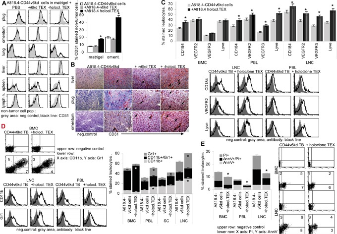
A818.4-CD44v6kd cells (5×10⁶) were embedded in matrigel and injected i.p. or were s.c. injected. Mice received 100μl NaCl or 100μg A818.4 holoclone TEX (i.p. or i.v.) 2x / week. A. Flow cytometry analysis of CD31+ cells in the matrigel plug, omentum, lung, liver, spleen and mesenteric lymph nodes, gating the non-tumor cells. The mean % stained non-tumor cells±SD (organs of 5 mice) and representative examples are shown. B. Immunohistochemistry of liver, plug and omentum stained with anti-CD31, staining of selected vessels is indicated by an arrow. C-E. Flow-cytometry analysis of (C) angiogenic factor receptor expression in BMC, PBL and LNC, (D) myeloid cells including myeloid-derived suppressor cells (CD11b+Gr1+) and (E) apoptotic cells in hematopoietic organs. (A, C-D) Mean values±SD of cells from 3-5 mice and representative examples are shown. Significant differences depending on holoclone TEX treatment are indicated: *. A818.4 holoclone TEX modulate host cells. This is demonstrated for (lymph)angiogenesis, myeloid and MDSC expansion and leukocyte apoptosis resistance.