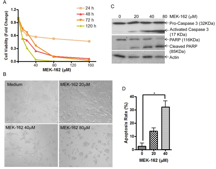
Figure 1. MEK-162 treatment inhibits murine pituitary corticotroph tumor cell proliferation in vitro.
A., Proliferation rates in murine pituitary corticotroph tumor AtT20 cells following MEK-162 treatment (10-160 μM) for 1 to 5 days were analyzed using CellTiter-Glo® luminescent cell viability assay. B., Microscopic changes of AtT20 cells following 48 h MEK-162 treatment showed cell shrinkage and apoptotic bodies. C., Caspase-3 and PARP cleavages, the hall markers of apoptosis, were detected by Western blotting following MEK-162 treatment for 48 h. D., The apoptotic induction rate of MEK-162 treatment for 48 h was detected using a caspase-3 colorimetric assay kit. Each bar indicates the mean ± standard error of triplicate tests. *p < 0.05. Data shown are representative of at least three independently performed experiments.

