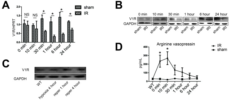
Figure 5. Activation of the vasopressin system during liver IR.
A., B., D. Liver samples were harvested from B6 mice that were either sham-operated (sham group) or subjected to 90 minutes of partial warm ischemia, followed by 0, 10, 30 minutes, 1, 6 or 24 hours of reperfusion (IR group, n = 3-6/group). A. The V1R gene expression was detected by real-time quantitative PCR, data were expressed as fold increase (mean±SD) above the sham group after 0 minute of reperfusion (set as 1). B. Western blot analysis of V1R expression in IR liver lobes. Densitometric values were shown in Figure S2A. C. Western blot analysis of V1R expression in isolated hepatocytes. Primary hepatocytes were subjected to hypoxia (SO2:3%) for 4 hours, hypoxia for 4 hours and restoration of oxygen supply (95% O2/5% CO2) for 1 (reper 1 hour) or 4 (reper 4 hour) hours. Primary hepatocytes from wild type B6 mice (WT) were subjected to normal oxygen supply (21% O2/5% CO2). Densitometric values of the western blot result were shown in Figure S2B. D. Serum concentrations of arginine vasopressin during hepatic IR. Data were expressed as mean±SD, *p < 0.05 as compared with sham group, NS, no significance. V1R, arginine vasopressin V1 receptor; PCR, polymerase chain reaction; IR, ischemia-reperfusion.

