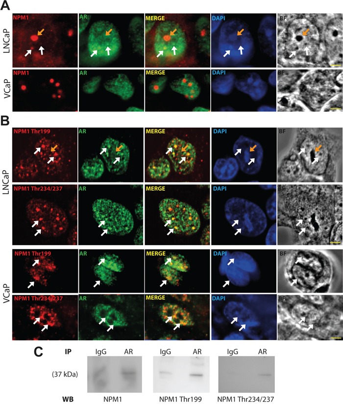
Figure 1. AR and Thr199 and Thr234/237 NPM1 localized in sub-nuclear structures.

A and B. LNCaP and VCaP cells were starved for 24 hours and then stimulated with 10 nmol/L DHT for 1 hour. Cells were fixed with methanol, immunostained (NPM1, AR and Thr199 and Thr234/237 NPM1) and analyzed by spinning disk fluorescence microscopy. Residual blurring was removed by spatial deconvolution. Nuclei were stained with DAPI. Scale bars, 2.5 μm. White arrows show the co-immunofluorescences and orange arrows show nucleoli. C. Nuclei of stimulated LNCaP were purified and AR was immuno-precipitated. The presence of NPM1 and its two phosphorylated forms were revealed by Western blotting.
