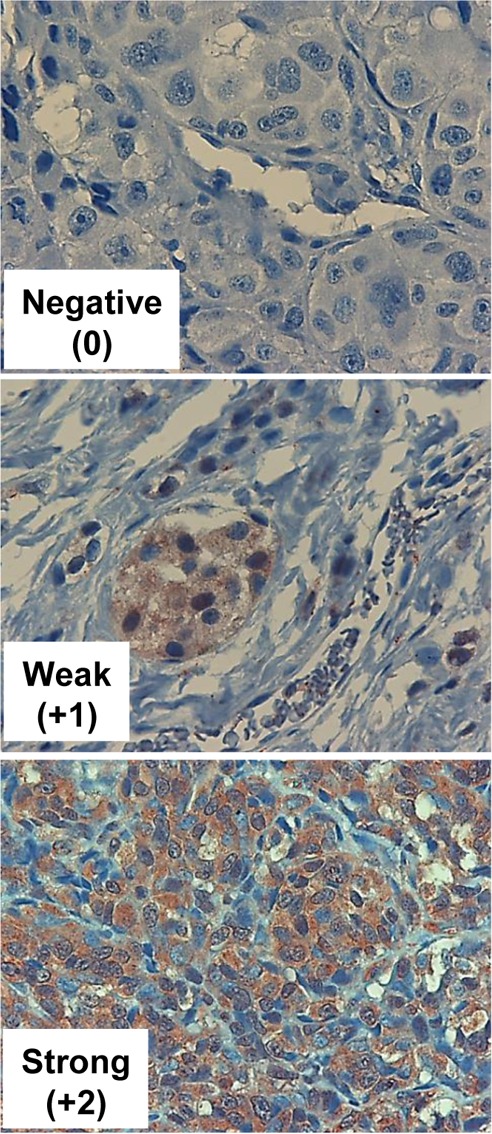
Figure 3. Hpa2 immunostaining.

Melanoma metastases were subjected to immunostaining applying anti-Hpa2 polyclonal antibody. Shown are representative photomicrographs of tumors that exhibit no (0, upper panel), weak (+1, middle panels) or strong (+2, lower panels) staining of Hpa2. Original magnification x100.
