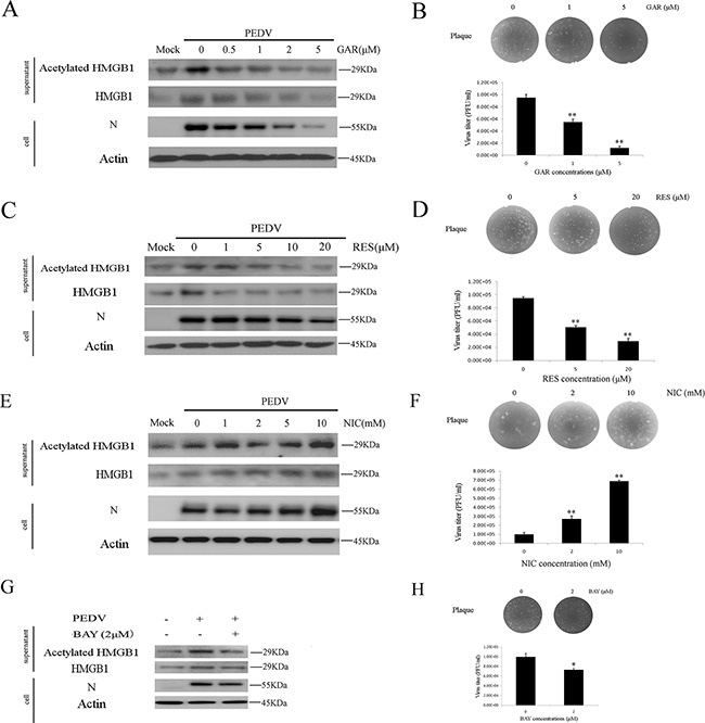
Figure 3. Histone acetyltransferase, SIRT1, and NF-κB are involved in HMGB1 acetylation.

The protein concentration in the supernatant was determined using the Bradford assay. An equal amount of proteins were used for western blot. A. Vero cells were treated with different concentrations GAR (HAT inhibitor) for 1h before infected with PEDV (MOI=0.1) in the presence of different concentrations of GAR for 24h. The acetylated HMGB1, total HMGB1 in the supernatant and PEDV-N in the cells were analyzed by western blot. B. The virus titer in the supernatant after GAR treatment was measured by the plaque formation assay. C. Vero cells were treated with different concentrations RES (SIRT1 activator) for 1h before infected with PEDV (MOI=0.1) in the presence of different concentrations of RES for 24h. The acetylated HMGB1 or total HMGB1 in the supernatant and PEDV-N in cells were analyzed by western blot. D. The virus titer in the supernatant after RES treatment was measured by the plaque formation assay. E. Vero cells were treated with different concentrations NIC (SIRT1 inhibitor) for 1h before infected with PEDV (MOI=0.1) in the presence of different concentrations of NIC for 24h. The acetylated HMGB1 or total HMGB1 in the supernatant and PEDV-N in cells were analyzed by western blot. F. The virus titer in the supernatant after NIC treatment was measured by plaque formation assay. G. Vero cells infected with PEDV (MOI=0.1) were treated with BAY (NF-κB inhibitor) for 24h. The acetylation/release of HMGB1 in the supernatant and PEDV-N in the cells were evaluated by western blot. H. The virus titer in the supernatant after BAY treatment was measured by the plaque formation assay. The results are the representative of at least two different experiments. The results represent the means ±SD of triplicate determinations. One-way ANOVA; *, P < 0.05; **, P < 0.01.
