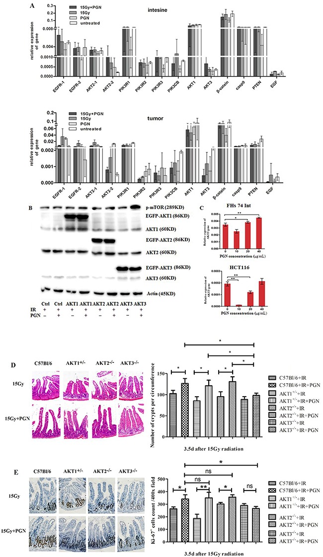
Figure 5. AKT3 was implicated in PGN's distinct effects on intestinal and tumor cell proliferation after IR.

A. EGFR (two transcripts), AKT2 (two transcripts), PIK3R1, PIK3R2, PIK3R3, β-catenin, AKT3, AKT1, Casp9, PTEN, PIK3CB, and EGF expression were detected by real-time PCR. Data are shown as the mean of 2(Ct, actin-Ct, target)±standard deviation. B. Western blots for AKT1/2/3 and p-mTOR in AKT1/2/3 overexpressing HCT116 cells. ‘EGFP-AKT' represents exogenously expressed AKT whereas ‘AKT’ represents endogenously expressed protein. β-actin was used as loading control. C. Real-time PCR analysis of AKT3 expression in FHS 74 Int and HCT116 cells following treatment with 0, 10, 20, and 40 μg/mL PGN (*, p<0.05; **, p<0.01). D. H&E staining of the cross section intestines of AKT1+/−, AKT2−/−, AKT3−/−, and control C57Bl/6 mice, irradiated or treated with PGN after IR. The crypts per circumference were counted. PGN had no effect on the number of crypts in irradiated AKT3−/− mice. E. Ki67 immunohistochemical staining showed that the number of Ki67+ crypt did not increase when irradiated AKT3−/− mice were treated with PGN. n=6 in each group; magnification: 400×. *, p <0.05; **, p <0.01.
