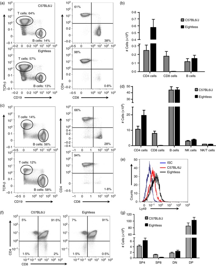
Figure 1.

Diminished CD8+ T cells in eightless mutant mice. (a). Flow cytometry analysis of the expression of CD19 versus T cell receptor‐β (TCR‐β) by cells from two inguinal lymph nodes (left panels), and CD8 versus CD4 (right panels, gated on TCR‐β + cells), from wild‐type or eightless mice. Numbers in quadrants indicate per cent cells in each. (b) Graph presenting absolute numbers of CD4 T, CD8 T and B cells in the spleen of wild‐type and eightless mice. (c) Same as in (a) using cells from the spleen. (d) Absolute numbers of CD4 T, CD8 T, natural killer (NK), NKT and B cells in the spleens of wild‐type and eightless mice. (e) Histogram plot of Ly49 expression level on NK cells (gated on NK1.1+ TCR‐β − cells) from wild‐type (red line) or eightless (black line) mice. ISC‐isotype control (blue line). (f) Flow cytometry analysis of the expression of CD8 versus CD4 by thymocytes from wild‐type or eightless mice. (g) Absolute numbers of CD4 and CD8 T double‐negative, double‐positive and either CD4 or CD8 single‐positive thymocytes in the thymus of wild‐type and eightless mice. (n = 6 mice, a two tailed unpaired t‐test was used for statistical analysis). Scale bars represent ± SEM. [Colour figure can be viewed at wileyonlinelibrary.com]
