Erratum
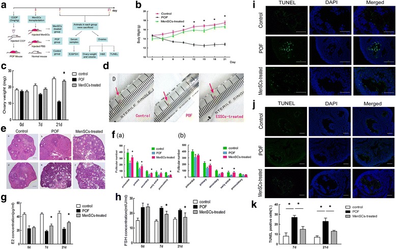
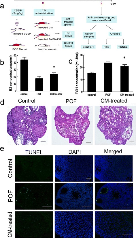
The original article [1] contains errors in Figs. 3i, j and 5e whereby the first column of each sub-panel is incorrectly labelled as having used GFP-staining; instead, the images were generated using TUNEL assays.
Fig. 3.

MenSC transplantation improves ovarian function after chemotherapy-induced injury. a Schematic of the experimental procedure used to explore the reparative effects of MenSCs in POF mice. b Changes in body weight between three groups (data expressed as mean ± SEM, *P < 0.05). c Changes in ovary weight across the three groups after 7 and 21 days (data expressed as mean ± SEM, *P < 0.05). d Macroscopic ovarian ovarian sizes in the three groups after 21 days. e Representative images showing H & E-stained ovary tissue sections in each group after 7 and 21 days. Scale bars = 100 μm. f Changes in follicle numbers in the three groups at 7 days (a) and 21 days (b) after MenSC transplantation (data expressed as mean ± SEM, *P < 0.05). g Serum E2 levels measured in each of the three groups. h Serum FSH levels measured in each of the three groups (data expressed as mean ± SEM, *P < 0.05). i Representative photograph showing TUNEL staining in ovary tissue sections after 7 days in each of the three groups. j Photograph showing TUNEL staining in ovary tissue sections after 21 days in each of the three groups. TUNEL-positive cells labelled green, and nuclei labelled blue (DAPI). Scale bars = 200 μm. k Quantitative analysis showing the percentage of TUNEL-positive cells in each group at 7 and 21 days after treatment (data expressed as mean ± SEM, *P < 0.05). CDDP cisplatin, DAPI 4′,6-diamidino-2-phenylindole, E2 oestradiol, FSH follicle-stimulating hormone, H&E haematoxylin and eosin, MenSC menstrual-derived stem cell, PBS phosphate-buffered saline, POF premature ovarian failure, TUNEL terminal deoxynucleotidyl transferase mediated dUTP nick end labelling
Fig. 5.

CM obtained from MenSCs improve ovarian function following chemotherapy-induced injury. a Schematic of the experimental procedure used to explore the reparative effects of CM in POF mice. b Serum E2 levels were measured in each of the three groups after 7 days. c Serum FSH levels were measured in each of the three groups after 7 days (data expressed as mean ± SEM, *P < 0.05). d Representative photomicrograph showing the results of H&E staining in each group at 7 days after injury. Scale bars = 100 μm. e Apoptosis evaluated using TUNEL staining in each group. Scale bars = 200 μm. CM conditioned media, CDDP cisplatin, DAPI 4′,6-diamidino-2-phenylindole, E2 oestradiol, FSH follicle-stimulating hormone, H&E haematoxylin and eosin, POF premature ovarian failure, TUNEL terminal deoxynucleotidyl transferase mediated dUTP nick end labelling
Consequently, the correct version of each figure can be seen below in this erratum.
Footnotes
The online version of the original article can be found under doi:10.1186/s13287-016-0458-1.
Reference
- 1.Wang Z, et al. Study of the reparative effects of menstrual-derived stem cells on premature ovarian failure in mice. Curr Stem Cell Res Ther. 2017;8:11. doi: 10.1186/s13287-016-0458-1. [DOI] [PMC free article] [PubMed] [Google Scholar]


