

Abstract
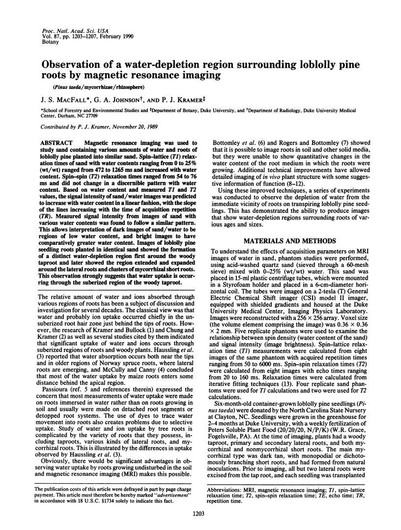
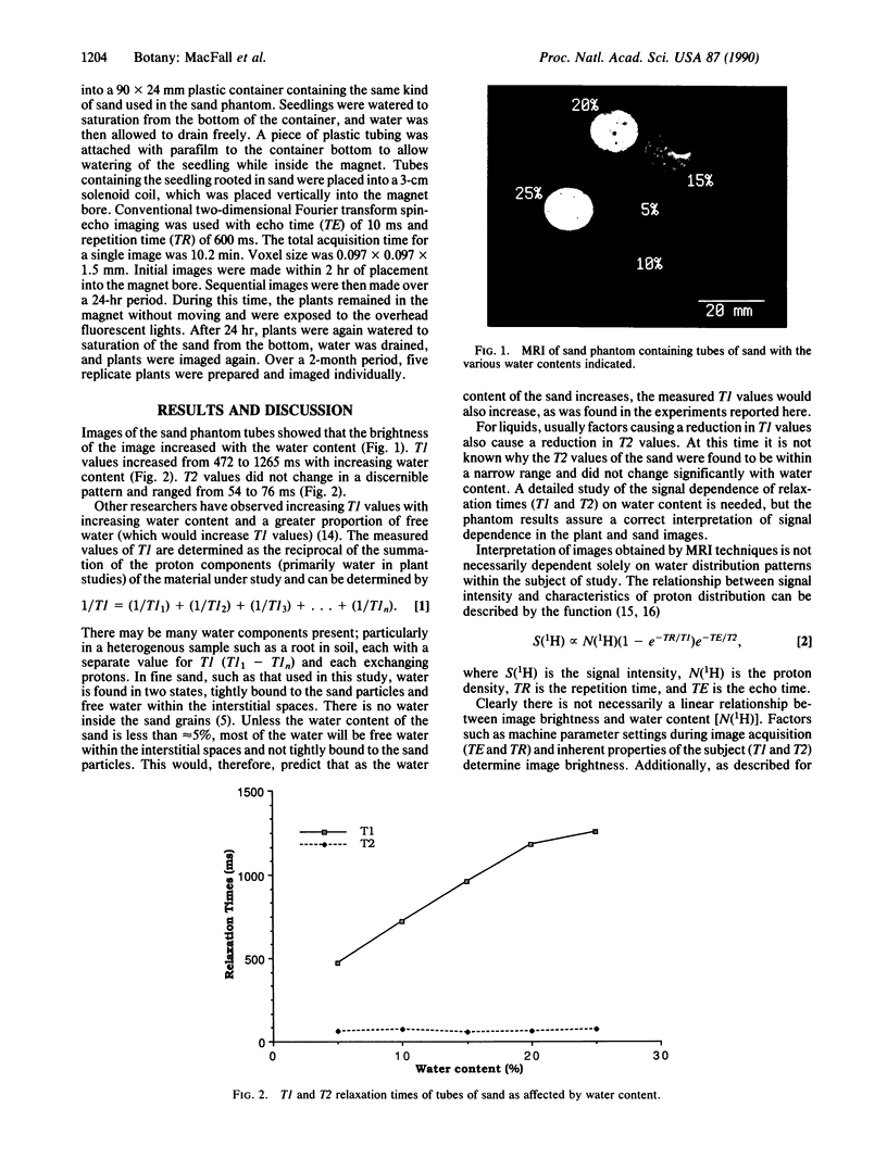
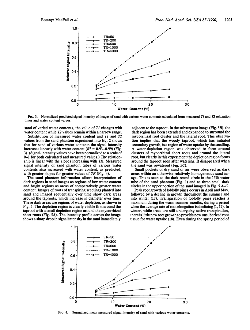
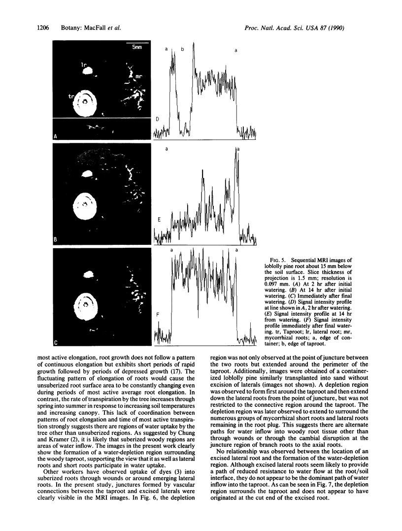
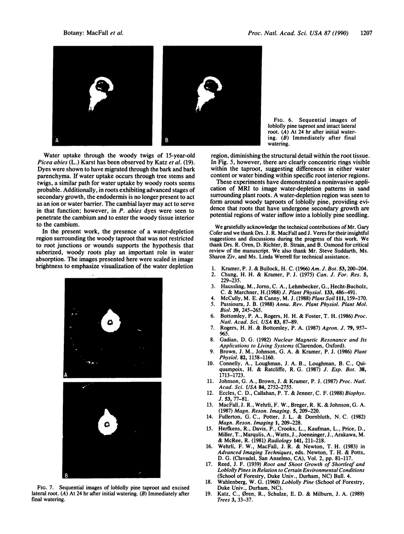
Magnetic resonance imaging was used to study sand containing various amounts of water and roots of loblolly pine planted into similar sand. Spin-lattice (T1) relaxation times of sand with water contents ranging from 0 to 25% (wt/wt) ranged from 472 to 1265 ms and increased with water content. Spin-spin (T2) relaxation times ranged from 54 to 76 ms and did not change in a discernible pattern with water content. Based on water content and measured T1 and T2 values, the signal intensity of sand/water images was predicted to increase with water content in a linear fashion, with the slope of the lines increasing with the time of acquisition repetition (TR). Measured signal intensity from images of sand with various water contents was found to follow a similar pattern. This allows interpretation of dark images of sand/water to be regions of low water content, and bright images to have comparatively greater water content. Images of loblolly pine seedling roots planted in identical sand showed the formation of a distinct water-depletion region first around the woody taproot and later showed the region extended and expanded around the lateral roots and clusters of mycorrhizal short roots. This observation strongly suggests that water uptake is occurring through the suberized region of the woody taproot.
Full text
PDF
Images in this article
Selected References
These references are in PubMed. This may not be the complete list of references from this article.
- Bottomley P. A., Rogers H. H., Foster T. H. NMR imaging shows water distribution and transport in plant root systems in situ. Proc Natl Acad Sci U S A. 1986 Jan;83(1):87–89. doi: 10.1073/pnas.83.1.87. [DOI] [PMC free article] [PubMed] [Google Scholar]
- Brown J. M., Johnson G. A., Kramer P. J. In Vivo Magnetic Resonance Microscopy of Changing Water Content in Pelargonium hortorum Roots. Plant Physiol. 1986 Dec;82(4):1158–1160. doi: 10.1104/pp.82.4.1158. [DOI] [PMC free article] [PubMed] [Google Scholar]
- Eccles C. D., Callaghan P. T., Jenner C. F. Measurement of the self-diffusion coefficient of water as a function of position in wheat grain using nuclear magnetic resonance imaging. Biophys J. 1988 Jan;53(1):77–81. doi: 10.1016/S0006-3495(88)83067-4. [DOI] [PMC free article] [PubMed] [Google Scholar]
- Fullerton G. D., Potter J. L., Dornbluth N. C. NMR relaxation of protons in tissues and other macromolecular water solutions. Magn Reson Imaging. 1982;1(4):209–226. doi: 10.1016/0730-725x(82)90172-2. [DOI] [PubMed] [Google Scholar]
- Herfkens R., Davis P., Crooks L., Kaufman L., Price D., Miller T., Margulis A. R., Watts J., Hoenninger J., Arakawa M. Nuclear magnetic resonance imaging of the abnormal live rat and correlations with tissue characteristics. Radiology. 1981 Oct;141(1):211–218. doi: 10.1148/radiology.141.1.7197379. [DOI] [PubMed] [Google Scholar]
- Johnson G. A., Brown J., Kramer P. J. Magnetic resonance microscopy of changes in water content in stems of transpiring plants. Proc Natl Acad Sci U S A. 1987 May;84(9):2752–2755. doi: 10.1073/pnas.84.9.2752. [DOI] [PMC free article] [PubMed] [Google Scholar]
- MacFall J. R., Wehrli F. W., Breger R. K., Johnson G. A. Methodology for the measurement and analysis of relaxation times in proton imaging. Magn Reson Imaging. 1987;5(3):209–220. doi: 10.1016/0730-725x(87)90022-1. [DOI] [PubMed] [Google Scholar]
- Turkki P. R., Ingerman L., Schroeder L. A., Chung R. S., Chen M., Dearlove J. Plasma pyridoxal phosphate as indicator of vitamin B6 status in morbidly obese women after gastric restriction surgery. Nutrition. 1989 Jul-Aug;5(4):229–235. [PubMed] [Google Scholar]