Abstract
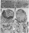
An unusual type of virus has been isolated from larvae of the cabbage looper, Trichoplusia ni (Lepidoptera; Noctuidae). The virus infects a variety of tissues, including fat body, epidermis, and tracheal matrix, causing a chronic, fatal disease. Viral replication begins in the nucleus and is accompanied by invagination of the nuclear envelope and extensive nuclear and cellular hypertrophy. The nuclear envelope eventually ruptures and fragments, after which viral-induced membranes are assembled along planes through the cell and around its periphery. Subsequently, these membranes coalesce, partitioning most of the cell, including viroplasms and virions in various stages of assembly, among a cluster of vesicles. The vesicles dissociate and are liberated into the hemolymph where they accumulate in large numbers (>108 vesicles per ml), causing the blood to become opaque white. The virus has been isolated from T. ni and transmitted per os and by injection to T. ni and several other species of the family Noctuidae. The virions produced by this virus are large (ca. 130 × 400 nm), enveloped, and allantoid in shape with complex symmetry and contain apparently linear, double-stranded DNA of Mr of ≈ 1.00 × 10⁸. The envelope contains subunits arranged in a hexagonal pattern that impart a distinctive reticular appearance to virions in negatively stained preparations. The unique structural and developmental properties of this virus indicate that it is a member of a group of enveloped, double-stranded DNA viruses not observed previously.
Keywords: viral vesicles, cell cleavage, mitochondria, membrane synthesis, virion assembly
Full text
PDF


Images in this article
Selected References
These references are in PubMed. This may not be the complete list of references from this article.
- Classification and nomenclature of viruses. Fourth report of the International Committee on Taxonomy of Viruses. Intervirology. 1982;17(1-3):1–199. doi: 10.1159/000149278. [DOI] [PubMed] [Google Scholar]
- Harrap K. A. The structure of nuclear polyhedrosis viruses. II. The virus particle. Virology. 1972 Oct;50(1):124–132. doi: 10.1016/0042-6822(72)90352-2. [DOI] [PubMed] [Google Scholar]
- Kelly D. C., Vance D. E. The lipid content of two iridescent viruses. J Gen Virol. 1973 Nov;21(2):417–423. doi: 10.1099/0022-1317-21-2-417. [DOI] [PubMed] [Google Scholar]
- Miller L. K., Dawes K. P. Restriction endonuclease analysis to distinguish two closely related nuclear polyhedrosis viruses: Autographa californica MNPV and Trichoplusia ni MNPV. Appl Environ Microbiol. 1978 Jun;35(6):1206–1210. doi: 10.1128/aem.35.6.1206-1210.1978. [DOI] [PMC free article] [PubMed] [Google Scholar]
- Stoltz A. B. The structure of icosahedral cytoplasmic deoxyriboviruses. II. An alternative model. J Ultrastruct Res. 1973 Apr;43(1):58–74. doi: 10.1016/s0022-5320(73)90070-1. [DOI] [PubMed] [Google Scholar]
- Wrigley N. G. An electron microscope study of the structure of Sericesthis iridescent virus. J Gen Virol. 1969 Jul;5(1):123–134. doi: 10.1099/0022-1317-5-1-123. [DOI] [PubMed] [Google Scholar]


