ABSTRACT
Objective:
To evaluate mouthpiece ventilation (MPV) in patients with Duchenne muscular dystrophy (DMD) who are noncompliant with noninvasive ventilation (NIV).
Methods:
We evaluated four young patients with DMD who had previously refused to undergo NIV. Each patient was reassessed and encouraged to try MPV.
Results:
The four patients tolerated MPV well and were compliant with NIV at home. MPV proved to be preferable and more comfortable than NIV with any other type of interface. Two of the patients required overnight NIV and eventually agreed to use a nasal mask during the night.
Conclusions:
The advantages of MPV over other types of NIV include fewer speech problems, better appearance, and less impact on the patient, eliminating the risk of skin breakdown, gastric distension, conjunctivitis, and claustrophobia. The use of a mouthpiece interface should be always considered in patients with DMD who need to start NIV, in order to promote a positive approach and a rapid acceptance of NIV. Using MPV during the daytime makes patients feel safe and more likely to use NIV at night. In addition, MPV increases treatment compliance for those who refuse to use other types of interfaces.
Keywords: Muscular dystrophy, Duchenne; Noninvasive ventilation; Patient compliance
RESUMO
Objetivo:
Avaliar a ventilação bucal (VB) em pacientes com distrofia muscular de Duchenne (DMD) não aderentes à ventilação não invasiva (VNI).
Métodos:
Foram avaliados quatro pacientes jovens com DMD que anteriormente recusaram-se a se submeter à VNI. Cada paciente foi reavaliado e encorajado a tentar VB.
Resultados:
Os quatro pacientes toleraram bem a VB e aderiram ao uso de VNI em casa. O uso de VB provou ser uma alternativa preferível e mais confortável que o uso de VNI com qualquer outro tipo de interface. Dois dos pacientes necessitaram de VNI noturna e eventualmente aceitaram utilizar uma máscara nasal durante a noite.
Conclusões:
As vantagens da VB sobre outros tipos de VNI incluem menores problemas na fala, melhor aparência e menor impacto no paciente, eliminando o risco de lesões na pele, distensão gástrica, conjuntivite e claustrofobia. O uso da interface bucal sempre deve ser considerado em pacientes com DMD que necessitam iniciar VNI a fim de promover uma abordagem positiva e uma rápida aceitação da VNI. O uso diurno de VB faz com que os pacientes sintam-se seguros e mais propensos a utilizar VNI à noite. Além disso, a VB aumenta a adesão ao tratamento naqueles pacientes que se recusam a utilizar outros tipos de interfaces.
INTRODUCTION
Duchenne muscular dystrophy (DMD) is one of the most common neuromuscular diseases in childhood. The young patient often needs noninvasive mechanical ventilation (NIV) due to the development of respiratory failure, reduction in FVC, or nocturnal hypoventilation symptoms. 1 - 3 Patients with neuromuscular disease might also function extremely well with minimal symptoms despite significant reductions in FVC and severe nocturnal oxygen desaturation. 4 For this reason and for the poor tolerance of the interface, sometimes the young patient does not accept NIV easily. Poor tolerance of the interface can be caused by various factors, such as excessive pressure of the mask on the face, excessive oral air leakage, anxiety of the patient because he or she is unable to call a family member, claustrophobia, and patient-ventilator dyssynchrony. 5 Thus, the interface plays a crucial role in tolerance and effectiveness of NIV use. Interfaces that cover the nose or the nose and mouth (oronasal interface) are the most commonly used; however, they are more likely to cause skin breakdown, gastric distension, conjunctivitis, and claustrophobia. 6 Sometimes the presence of deep cutaneous lesions precludes the use of NIV. The NIV interface, however, should be comfortable and reasonably airtight. Although there are various types of interfaces nowadays, the physiognomy of the patient often makes the choice of a suitable interface difficult. The most common drawbacks can be avoided by using mouthpiece NIV (MPV) interfaces, and, in recent years, MPV modes have been introduced to some of the portable ventilators available.
METHODS
The Monaldi Hospital Review Board approved the study. Patients were studied in our respiratory department between January of 2015 and April of 2015. We evaluated four young patients with DMD, with a mean of 18.5 years of age (Table 1). Three patients (labeled as patients 1, 2, and 3) showed reductions in FVC (< 50% than the previous control measurement) and progressive decreases in PaO2. The four patients presented with an FVC ≤ 1 L, and patients 1 and 4 presented with hypercapnia and nocturnal respiratory failure. Patient 1 presented with obstructive sleep-disordered breathing (SDB), and sleep monitoring demonstrated an apnea-hypopnea index = 20 events/h. Patient 4 presented with sleep hypoventilation syndrome; sleep monitoring demonstrated an increase in PaCO2 > 10 mmHg at waking in the morning (when compared with awake supine values) and sustained hypoxemia not related to apnea or hypopnea (SaO2 < 90% over 30% of the monitoring period during sleep; T90 > 30%), demonstrating SDB.
Table 1. Characteristics of the patients and noninvasive ventilation settings.
| Patient | Age, years | FVC, L | PaO2 | PaCO2 | SDB | Cause of NIV refusal | Mode | IPAP, cmH2O | EPAP, cmH2O | IT, s | RT, s | RR, cycles/min |
|---|---|---|---|---|---|---|---|---|---|---|---|---|
| 1 | 21 | 1,07 | 89 | 47 | Yes | Fear and anxiety | MPV/PC | 12 | 0 | 1,2 | 3 | 0 |
| 2 | 16 | 0,91 | 69 | 45 | No | Claustrophobia | MPV/PC | 14 | 0 | 1,2 | 3 | 0 |
| 3 | 18 | 0,96 | 70 | 44 | No | Skin intolerance | MPV/PC | 14 | 0 | 1,2 | 3 | 0 |
| 4 | 19 | 1,02 | 81 | 51 | Yes | Fear and anxiety | MPV/PC | 15 | 0 | 1,4 | 4 | 0 |
SDB: sleep-disordered breathing; NIV: noninvasive ventilation; IPAP: inspiratory positive airway pressure; EPAP: expiratory positive airway pressure; IT: inspiratory time; RT: rise time; RR: respiratory rate; and MPV/PC: mouthpiece ventilation/pressure control.
The patients had previously refused to use NIV due to claustrophobia, fear of being unable to communicate with family members while using it, and annoying leaks. Each patient was reassessed and proposed to try using MPV. All patients initiating NIV/MPV were treated in a day hospital. No patient was excluded from MPV as a result of bulbar impairment.
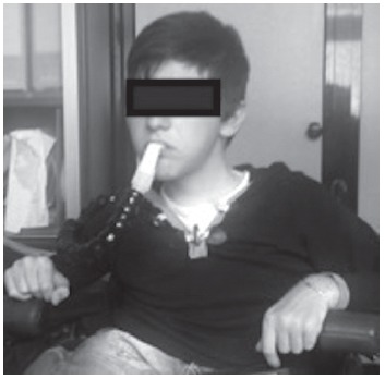
The subjects were submitted to NIV using a mechanical ventilator (Trilogy; Philips Respironics, Murrysville, PA, USA) that has a dedicated particular function designated "kiss trigger" MPV and an arm dedicated to support the mouthpiece in order to facilitate its use and allow the patient to stay on a wheelchair (Figure 1). During the adaptation phase, we presented the mouthpiece interface to the patients and selected the most comfortable position of the support arm according to the postural impairment of the patient. The preferred mode in the initial phase was pressure control, with a support of 8-10 cmH2O, which was gradually increased until a suitable tidal volume, an optimal SpO2 (confirmed by blood gas analysis), a stable HR, and good chest expansion (measured by electrical impedance tomography-PulmoVista 500; Dräger Medical GmbH, Lübeck, Germany) were reached. Inspiratory time was set at 1.2 s (for patients 1, 2, and 3), and 1.4 s (for patient 4). For all patients, the expiratory positive airway pressure (EPAP) was set to 0 cmH2O; the rise time amounted to 3 s (for patients 1, 2, and 3) and 4 s (for patient 4). Respiratory rate (RR) was set to 0 breaths/min. Time of disconnection alarm was set at 15 min (Table 1). All patients tolerated this new ventilation mode well, with good compliance to the interface. All patients accepted the treatment, and they continued NIV at home. At this writing, none of the DMD patients had been unable to retain the mouthpiece as a result of weakness.
Figure 1. A patient during mouthpiece ventilation.

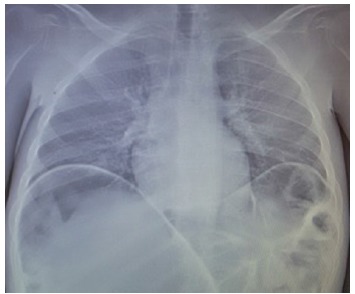
In our practice, patients are submitted to chest X-rays prior to treatment. The results for patients 2 and 3 showed dilated bowel loops and upward displacement of the diaphragm (Figure 2).
Figure 2. A chest X-ray showing dilated bowel loops and upward displacement of the diaphragm.

After an initial phase of adaptation to MPV during daytime hours, thanks to a greater feeling of security, patients 1 and 4 also accepted NIV with a nasal mask during the night. Patient 1 was adapted to spontaneous-timed average volume assured pressure support (ST-AVAPS) ventilation mode; maximum inspiratory positive airway pressure (IPAP) = 14 and 10 cmH2O; minimum IPAP = 10 cmH2O; EPAP = 6 cmH2O; AVAPS rate = 1 cmH2O/min; tidal volume = 650 mL; trigger flow = 4.0 L; cycle = 25%; rise time = 3 s; RR = 10 breaths/min; and inspiratory time = 1.2 s in a simple circuit configuration. Patient 4 was adapted to assist-control ventilation (ACV) mode; VC = 750 mL; RR = 12 breaths/min; inspiratory time = 1.2 s; decelerated flow; and EPAP = 4 cmH2O (Table 2).
Table 2. Mouthpiece ventilation settings for patients 1 and 2.
| Patient | Mode | IPAPmin, cmH2O | IPAPmax, cmH2O | EPAP, cmH2O | AVAPS rate | VT, L | TG | Cycle | RT, s | RR, breaths/min | IT, s | Interface |
|---|---|---|---|---|---|---|---|---|---|---|---|---|
| 1 | ST-AVAPS | 10 | 14 | 6 | 1 | 650 | 4 | 25% | 3 | 10 | 1,2 | Nasal |
| 4 | AC | 4 | 750 | 1,2 | Nasal |
IPAPmin: minimum inspiratory positive airway pressure; IPAPmax: maximum inspiratory positive airway pressure; EPAP: expiratory positive airway pressure; AVAPS: average volume assured pressure support; VT: tidal volume; TG: trigger flow; RT: rise time; RR: respiratory rate; IT: inspiratory time; ST: spontaneous-timed; and AC: assist-control
Oxyhemoglobin saturation was measured using pulse oximetry (PalmSAT 2500A; Nonin Medical, Plymouth, MN, USA), and PaCO2 was measured using a transcutaneous blood gas monitor (Linde MicroGas 7650; Linde Medical Sensors AG, Basel, Switzerland) or arterial blood gases when the transcutaneous monitor was unavailable while the patient was awake. Adequate ventilatory support was assessed using a combination of clinical assessment, normal overnight oximetry, serial daytime measures of PaCO2, and downloaded data from the devices.
RESULTS
All of the patients had previously refused to use NIV, but accepted treatment with MPV. The preferred mode was pressure control (Table 1). IPAP was set between 10 and 14 cmH2O, which ensured an optimal tidal volume (8-10 mL/kg). No back-up rate was needed during daytime, so no air blew into the face of the patients. In this configuration, the equipment could be set without EPAP. Alarms for apnea, minimum pressure, and minimum volume could be easily shut off in order to avoid their unnecessary activation. In most home volume-controlled ventilators, the minimum pressure alarm cannot be deactivated; therefore, it is necessary to set up positive end-expiratory pressure (often at 2 cmH2O), which, thanks to the resistance to the airflow created from the angle of the mouthpiece, creates a pressure that prevents the continuous activation of the alarms.
Patient 1 presented with sleep apnea syndrome. During night time, he was treated using ST-AVAPS mode, which allowed greater comfort and better fit. The use of EPAP = 6 cmH2O helped to reduce apnea. A brief expiratory cycle was more physiological. A trigger with an average sensitivity was better tolerated than a more sensitive trigger, which was initially set. The patient had good peak inspiratory flow and a great number of triggered cycles. Patient 2 presented with sleep hypoventilation syndrome and adapted to the ACV mode well, reducing respiratory effort and improving nocturnal breathing pattern.
DISCUSSION
Respiratory disease is a nearly inevitable complication in patients with DMD and represents the underlying cause of death in 70% of the patients with less than 25 years of age. 7 It has been widely demonstrated that mechanical ventilation corrects respiratory failure and can prolong the life of those patients. 8 It is known that NIV improves gas exchange, relieves shortness of breath, allows inspiratory muscles to rest, and reduces the incidence of nosocomial infections. In addition, mortality and hospitalizations due to respiratory failure decrease. 9 In our experience, the selection of an appropriate interface is crucial for successful NIV. Nasal and oronasal masks are the most practical (and most commonly used) for NIV. 10 The main limitations to the use of these interfaces are claustrophobia, discomfort, and skin lesions. 11 , 12 The application of an oronasal interface can worsen social interaction, since it impairs eating, drinking, and talking. This type of mask changes the patient's perception of himself and may have negative psychological consequences. 13 The preferable and more comfortable alternative is MPV via a 15- or 22-mm mouthpiece device; however, more active participation of the patient is necessary than with the use of traditional masks. We preferred the ''kiss trigger'' MPV function of the ventilator, because the patient has only to touch it for air delivery. Patients easily trigger the ventilator with mouth pressure. The open-circuit MPV is safe and comfortable to patients confined to wheelchairs. There is a system in which the mouthpiece should be placed close to the mouth by the adjustable arm, and the patient can grab the mouthpiece as desired. It should be removable for talking, eating, or breathing independently. The most significant advantages of MPV when compared with the use of a nasal or an oronasal mask are that the mouthpiece interferes with speech to a lesser degree, improves appearance, reduces the negative impact on the patient, and eliminates the risk of skin breakdown and claustrophobia. 14 Additionally, it is safer than the other interfaces since it permits the use of glossopharyngeal breathing in the event of a sudden ventilator failure or an accidental disconnection from the ventilator.
A recent comparative study on the performance of different home ventilators showed that MPV and the use of a whisper valve were associated with a reduction in the number of alarms when compared with the other configurations available in the Trilogy (Philips Respironics) ventilator. The authors emphasized that the "kiss trigger" function in that ventilator is not comparable with functions in classical ventilators, since it is based on a flow signal technology that detects an alteration in the flow-by mode generated by the ventilator due to any reason, such as a partial obstruction via the mouth connection. 15
All of the patients in the present study had previously rejected the application of NIV due to the tightness of the interface and claustrophobia, resulting in poor compliance. The patients who required overnight NIV subsequently accepted the treatment via a nasal mask during the night hours. This was probably due to the fact that the use of MPV during daytime hours made the patients feel safe, gradually being confident enough for using NIV at night. The use of the nasal mask and MPV with that specific ventilator (Trilogy; Philips Respironics) allowed us to treat the patients who had previously refused nasal, oral, or oronasal interfaces.
The use of a mouthpiece interface should be always considered for patients with DMD who need to start NIV; it is useful to promote a positive approach and a rapid acceptance of the new condition. As time goes by, patients with DMD develop constant hypoventilation and need respiratory support 24 h a day; MPV can be valuable, particularly in patients who undergo NIV various hours a day and who present with skin lesions, gastric distension, or eye irritation. The use of a nasal or an oronasal mask may be alternated as well.
Footnotes
Financial support: None.
Study carried out in the Dipartimento di Fisiopatologia Respiratoria, Ospedale Monaldi di Napoli, Napoli, Italia.
REFERENCES
- 1.Hamada S, Ishikawa Y, Aoyagi T, Ishikawa Y, Minami R, Bach JR. Indicators for ventilator use in Duchenne muscular dystrophy. Respir Med. 2011;105(4):625–629. doi: 10.1016/j.rmed.2010.12.005. http://dx.doi.org/10.1016/j.rmed.2010.12.005 [DOI] [PubMed] [Google Scholar]
- 2.Toussaint M, Chatwin M, Soudon P. Mechanical ventilation in Duchenne patients with chronic respiratory insufficiency clinical implications of 20 years published experience. Chron Respir Dis. 2007;4(3):167–177. doi: 10.1177/1479972307080697. http://dx.doi.org/10.1177/1479972307080697 [DOI] [PubMed] [Google Scholar]
- 3.Nardi J, Prigent H, Garnier B, Lebargy F, Quera-Salva MA, Orlikowski D. Efficiency of invasive mechanical ventilation during sleep in Duchenne muscular dystrophy. Sleep Med. 2012;13(8):1056–1065. doi: 10.1016/j.sleep.2012.05.014. http://dx.doi.org/10.1016/j.sleep.2012.05.014 [DOI] [PubMed] [Google Scholar]
- 4.Jeppesen J, Green A, Steffensen BF, Rahbek J. The Duchenne muscular dystrophy population in. Denmark, 1977- 2001:prevalence–prevalence. doi: 10.1016/s0960-8966(03)00162-7. http://dx.doi.org/10.1016/S0960-8966(03)00162-7 [DOI] [PubMed] [Google Scholar]
- 5.Garuti G, Nicolini A, Grecchi B, Lusuardi M, Winck JC, Bach JR. Open circuit mouthpiece ventilation Concise clinical review. Rev Port Pneumol. 2014;20(4):211–218. doi: 10.1016/j.rppneu.2014.03.004. http://dx.doi.org/10.1016/j.rppneu.2014.03.004 [DOI] [PubMed] [Google Scholar]
- 6.Pisani L, Carlucci A, Nava S. Interfaces for noninvasive ventilation technical aspects and efficiency. Minerva Anestesiol. 2012;78(10):1154–1161. [PubMed] [Google Scholar]
- 7.Bach JR, Martinez D. Duchenne muscular dystrophy continuous noninvasive ventilatory support prolongs survival. Respir Care. 2011;56(6):744–750. doi: 10.4187/respcare.00831. http://dx.doi.org/10.4187/respcare.00831 [DOI] [PubMed] [Google Scholar]
- 8.Finder JD, Birnkrant D, Carl J, Farber HJ, Gozal D, Iannaccone ST. Respiratory care of the patient with Duchenne muscular dystrophy ATS consensus statement. Am J Respir Crit Care Med. 2004;170(4):456–465. doi: 10.1164/rccm.200307-885ST. http://dx.doi.org/10.1164/rccm.200307-885ST [DOI] [PubMed] [Google Scholar]
- 9.Bach JR, Rajaraman R, Ballanger F, Tzeng AC, Ishikawa Y, Kulessa R. Neuromuscular ventilator insufficiency effect of home mechanical ventilator use vs oxygen therapy on pneumonia and hospitalization rates. Am J Phys Rehabil. 1998;77(1):8–19. doi: 10.1097/00002060-199801000-00003. http://dx.doi.org/10.1097/00002060-199801000-00003 [DOI] [PubMed] [Google Scholar]
- 10.Hess DR. Noninvasive ventilation in neuromuscular disease equipment and application. Respir Care. 2006;51(8):896–911. [PubMed] [Google Scholar]
- 11.Gregoretti C, Confalonieri M, Navalesi P, Squadrone V, Frigerio P, Beltrame F. Evaluation of patient skin breakdown and comfort with a new face mask for non-invasive ventilation a multi-center study. Intensive Care Med. 2002;28(3):278–284. doi: 10.1007/s00134-002-1208-7. http://dx.doi.org/10.1007/s00134-002-1208-7 [DOI] [PubMed] [Google Scholar]
- 12.Antón A, Tarrega J, Giner J, Güell R, Sanchis J. Acute physiologic effects of nasal and full-face masks during noninvasive positive-pressure ventilation in patients with acute exacerbations of chronic obstructive pulmonary disease. Respir Care. 2003;48(10):922–925. [PubMed] [Google Scholar]
- 13.Sferrazza Papa GF, Di Marco F, Akoumianaki E, Brochard L. Recent advances in interfaces for non-invasive ventilation from bench studies to practical issues. Minerva Anestesiol. 2012;78(10):1146–1153. [PubMed] [Google Scholar]
- 14.Benditt JO. Full-time noninvasive ventilation possible and desirable. Respir Care. 2006;51(9):1005–1012. [PubMed] [Google Scholar]
- 15.Khirani S, Ramirez A, Delord V, Leroux K, Lofaso F, Hautot S. Evaluation of ventilators for mouthpiece ventilation in neuromuscular disease. Respir Care. 2014;59(9):1329–1337. doi: 10.4187/respcare.03031. http://dx.doi.org/10.4187/respcare.03031 [DOI] [PubMed] [Google Scholar]


