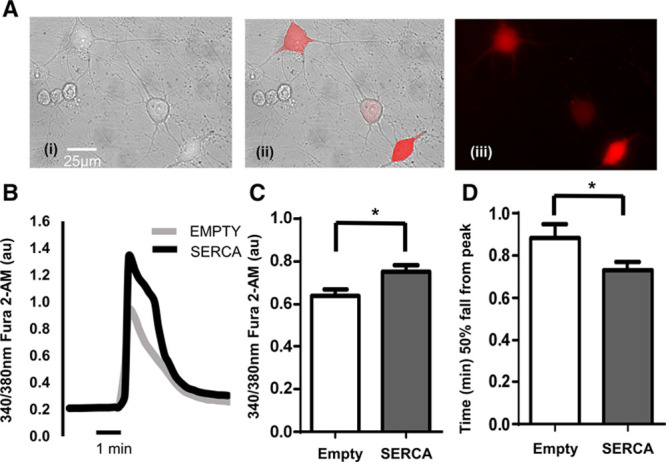
Figure 2.

A, Ad-mCherry-hATP2Aa transfected stellate ganglia neurons from (4 to 5 wk, 90–120 g) Sprague–Dawley (SD) rat. (i) Bright field image, (ii) composite, and (iii) excitation at 587 nm to excite mCherry fluorescent tag. Only cells expressing mCherry fluorescence were used for experiments. B, Example raw data trace from isolated stellate ganglia neurons of the young SD rat (gray line, Ad-mCherry [empty]; black line, Ad-mCherry-hATP2Aa [SERCA (sarcoendoplasmic reticulum calcium ATPase)]) exposed to 50 mmol/L of KCl (30 s) to depolarize the neuron resulting in an increase in intracellular free Ca2+ ([Ca2+]i). C, Group mean data showing peak depolarization-evoked intracellular free Ca2+ increase between Ad-mCherry (gray; n=57) and Ad-mCherry-hATP2Aa (black; n=68) transfected stellate neurons. D, Group mean data of 50% fall time of ([Ca2+]i) from the peak (Ad-mCherry, gray; n=37; Ad-mCherry-hATP2Aa, black; n=42). *P<0.05.
