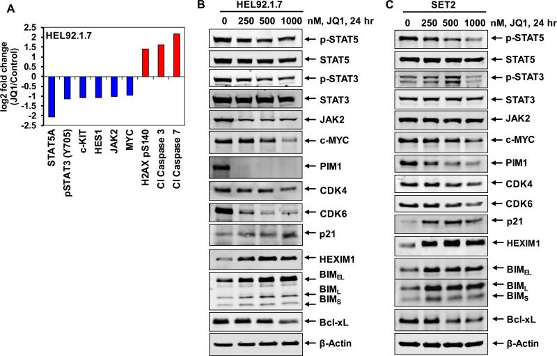
Figure 2. Treatment with JQ1 depletes the expression levels of pSTAT5, c-MYC, CDK4 and CDK6 with concomitant induction of HEXIM1 and BIM expression levels in sAML cells.
A. HEL92.1.7 cells were treated with JQ1 for 24 hours, and then analyzed by reverse phase protein analysis. Figure shows log2 fold change of selected targets in JQ1-treated cells relative to the untreated control cells. All fold changes shown are significant (p<0.05). B-C. HEL92.1.7 and SET-2 cells were treated with the indicated concentrations of JQ1 for 24 hours. Total cell lysates were prepared and immunoblot analyses were conducted as indicated. The expression levels of β-Actin in the lysates served as the loading control.

