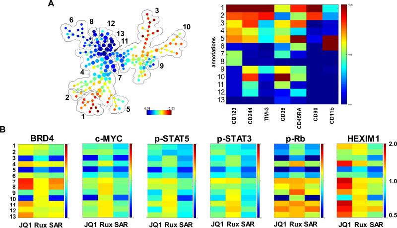
Figure 6. Treatment with JQ1 or JAK inhibitors ruxolitinib or SAR302503 depletes JAK/STAT signaling and attenuates c-MYC, BCL-2 and Bcl-xL protein expression in primary CD34+ secondary AML cells as determined by mass cytometry analysis.
Primary CD34+ secondary AML cells were treated with JQ1 or ruxolitinib or SAR302503 for 16 hours. At the end of treatment, cells were fixed and labeled with a cocktail of primary antibodies conjugated to rare metal elements. Mass cytometry ‘CyTOF’ analysis was conducted and data were normalized and exported. Data were further analyzed utilizing the SPADE software. A. SPADE tree colored by c-MYC and a heatmap of the 13 annotated clusters on the SPADE tree based on the expression levels of CD markers and cellular markers associated with stem cells or differentiated cells. B. Heatmaps of the protein expression changes caused by treatment with JQ1, ruxolitinib, or SAR302503 relative to the untreated control cells (set to 1) are shown.

