Abstract
Introduction:
Chromosomal microarray analysis (CMA) is recognized as the first-tier test in the genetic evaluation of children with developmental delays, intellectual disabilities, congenital anomalies and autism spectrum disorders of unknown etiology.
Array Design:
To optimize detection of clinically relevant copy number variants associated with these conditions, we designed a whole-genome microarray, FirstStepDx PLUS (FSDX). A set of 88,435 custom probes was added to the Affymetrix CytoScanHD platform targeting genomic regions strongly associated with these conditions. This combination of 2,784,985 total probes results in the highest probe coverage and clinical yield for these disorders.
Results and Discussion:
Clinical testing of this patient population is validated on DNA from either non-invasive buccal swabs or traditional blood samples. In this report we provide data demonstrating the analytic and clinical validity of FSDX and provide an overview of results from the first 7,570 consecutive patients tested clinically. We further demonstrate that buccal sampling is an effective method of obtaining DNA samples, which may provide improved results compared to traditional blood sampling for patients with neurodevelopmental disorders who exhibit somatic mosaicism.
Clinical Scenario
Neurodevelopmental disabilities, including developmental delays (DD), intellectual disabilities (ID), and autism spectrum disorders (ASD), affect up to 15% of children1. In the majority of cases, a child’s clinical presentation does not allow for a definitive etiological diagnosis. In such cases, CMA is recommended as the first-tier test that should be used to evaluate for a potential genetic etiology2 , 3 , 4 , 5 , 6 , 7. A definitive genetic diagnosis allows patients to more often receive appropriate medical care tailored to their condition, as reflected by medical management changes and improved access to necessary support and educational services8 , 9 , 10 , 11 , 12 , 13.
Test Description
FirstStepDx PLUS (FSDX) is an optimized clinical microarray test provided in the context of a comprehensive clinical service. Testing starts with either a non-invasive buccal swab sample or traditional blood sample from which DNA extraction using a Gentra Puregene® kit specific to the sample type (Qiagen, Inc., Valencia, CA) is performed in one of several contracted CLIA/CAP credentialed laboratories according to manufacturers’ protocols. High quality genomic DNA is fragmented, labeled and hybridized to FSDX arrays using reagents, equipment and methodology as specified by the manufacturer (Affymetrix, Inc., Santa Clara, CA)14. Washed arrays are scanned and raw data files are processed to CYCHP files using a reference file comprising at least 100 samples with normal array findings. Data analysis is performed using Chromosome Analysis Suite software version 2.0.1 (Affymetrix). Hybridization of patient DNA to oligonucleotide and SNP probes is independently compared against a previously analyzed cohort of normal samples to call CNVs and allele genotypes. The percentage mosaicism of whole-chromosome aneuploidies is determined using the average log2 ratio of the entire chromosome14.
Microarray Design
FSDX was optimized by the addition of 88,435 custom probes targeting genomic regions strongly associated with ID/DD/ASD15 , 16 , 17 , 18 , 19 , 20 , 21 , 22 , 23 , 24. This was effected, under GMP by Affymetrix, to the CytoScanHD platform using their custom microarray design process. This is consistent with the ACMG recommendation of “enrichment of probes targeting dosage-sensitive genes known to result in phenotypes consistent with common indications for a genomic screen”25. Critical regions that did not meet a desired probe density >1 probe/1000 bp on the CytoScanHD were supplemented with additional probe content to allow for improved detection of smaller deletions and duplications in these critical regions. Finally, additional probes were added to improve detection of CNVs encompassing genes involved in other well-characterized neurodevelopmental disorders, for example GAMT26 and GATM27. All incremental probes were added in substitution for probes deemed sub-optimal by Affymetrix and previously masked, bringing FSDX to a grand total of 2,784,985 probes. Custom SNP probes (n =416) on FSDX are targeted by 12 oligonucleotides, three for each strand of each allele, which is approximately double the typical probe coverage for SNPs.
Test Interpretation
CYCHP files are evaluated by ABMG certified cytogeneticists. Determination of CNVs is consistent with established cytogenetic standards. A minimum of 25-consecutive impacted probes is the baseline determinant for deletions and 50 probes for duplications independent of variant size. Rare CNVs are determined to be “pathogenic” if there is sufficient evidence published (at least two independent publications) to indicate that haploinsufficiency or triplosensitivity of the region or gene(s) involved is causative of clinical features or of sufficient overall size28. If, however, there is insufficient but at least preliminary evidence for a causative role for the region or gene(s) therein they are classified as variants of unknown significance (VOUS) independent of CNV size. Detection of these VOUS is important, as they serve as source for further delineation of causal variants as well as benign variants. (For a more complete discussion of this topic see the following link: http://blog.goldenhelix.com/clambert/sustaining-competitive-advantage-in-array-based-cytogenetics/.) Further, surveys of families who received reports containing VOUS findings indicate that these findings, when communicated appropriately, unquestionably contribute to families’ understanding of the disorder as well as their ability to explain it to others29 , 30 , 31.
Areas of absence of heterozygosity (AOH) are also classed as VOUS if of sufficient size and location to increase the risk for conditions with autosomal recessive inheritance or conditions with parent-of-origin/imprinting effects. Other CNVs are typically not reported after determination that they most likely represent normal common population variants and are contained in databases documenting presumptively benign CNVs, e.g., the Database of Genomic Variants (DGV)32. These parameters were standard independent of the microarray used for analysis in comparative studies.
Public Health Importance
A definitive genetic diagnosis facilitates patient access to appropriate and necessary medical and support services. Defining the underlying genetic cause of DD/ID/ASD and/or multiple congenital anomalies (MCA) in each unique patient is vital to understanding etiology, prognosis, and course. It informs physicians of potential comorbid conditions for which a patient should be evaluated and treated proactively and optimally. Improved understandings of the appropriate therapeutic and behavioral approaches to that patient are also enabled. Genetic testing is best provided in the context of an integrated service33, so FSDX aims to provide comprehensive, clear, readable, and personalized reports for the healthcare provider and a family-friendly section to facilitate understanding of the often-complex results. The report is complemented by availability of pre- and post-test genetic counseling and technical support to providers. Moreover, FSDX includes personalized insurance pre-authorization and appeals assistance intended to help overcome barriers encountered by both providers and families that, in many circumstances, prevent access to crucial genetic testing services10 , 11.
Published Reviews, Recommendations and Guidelines
The American College of Medical Genetics (ACMG)2 , 3, the American Academy of Child and Adolescent Psychiatry4 , 7, the American Academy of Pediatrics5, and the American Academy of Neurology/Child Neurology Society6 recommend CMA as the first-tier test in the genetic evaluation of children with unexplained DD, ID, or ASD. Considerable data supporting these guidelines are documented in numerous reviews and publications34 , 35 , 36 , 37 , 38 , 39. The ACMG has also published guidelines on both array design25 and the validation of arrays, including validation of a new version of a platform in use by the laboratory from the same manufacturer, and of additional sample types28.
EVIDENCE OVERVIEW
Validation of Novel (Blood vs. Buccal) Sample Type on the Established and Optimized Platforms
It is highly desirable to avail clinicians and families a less invasive sampling method for individuals with ID/DD/ASD due to the potential implications of venipuncture in many such individuals. We validated a buccal sampling methodology in conjunction with two independent CLIA-certified laboratories, ARUP (Salt Lake City, UT) and Fullerton Genetics Center (Asheville, NC) first on the CytoScanHD array and then the FSDX array. After preliminary consideration of multiple saliva and buccal DNA collection kits, we selected the ORAcollect-100® (DNA Genotek, Ottawa) (now cleared as a class II IVD medical device: ORAcollect-DX®) based upon ease of use for the intended patient population, ease of shipping processes, DNA stability, and post-extraction quality studies for further validation. Buccal swabs and blood samples from twenty-two individuals underwent parallel microarray analysis for concordance by each protocol. Twenty-two individuals’ buccal and blood samples were analyzed in terms of array quality control metrics and no significant difference between the two sample sources was observed. In addition, there was 100% concordance of CNV calls between the two sample types. Finally, twenty-three individuals’ blood and buccal samples underwent microarray analysis on the FSDX platform. Again no significant difference between the two sample sources in terms of quality metrics and 100% clinical concordance of CNV calls was observed.
Clinical Validation of a New Version of a Previously Established Platform
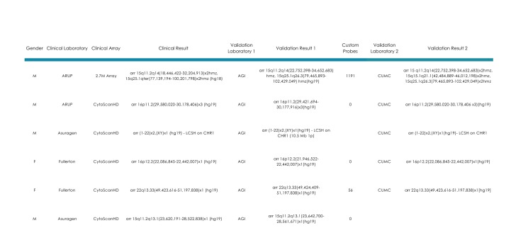
FSDX was validated independently in four CLIA-certified laboratories (Asuragen – Asuragen, Inc., Austin, TX; AGI – Affiliated Genetics, Inc., Salt Lake City, UT; Fullerton - Mission Hospital/Fullerton Genetics Center, Asheville, NC; CUMC – Columbia University Medical Center, Dept. of Pathology and Cell Biology, New York, NY) all previously familiar with performing CMA on CytoScanHD (or its predecessor the Affymetrix 2.7M Cytogenetics array) and cross-validated between these laboratories as well. Data demonstrating the concordance of FSDX with these alternative arrays and across laboratories are shown in Tables I-II.
Table I: Clinical validation of the FirstStepDx PLUS array Samples from patients analyzed clinically on commercially available Samples from patients analyzed clinically on commercially available Affymetrix microarrays were run independently on FSDX. Laboratory designations are as follows: Asuragen – Asuragen, Inc., Austin, TX; AGI – Affiliated Genetics, Inc., Salt Lake City, UT; Fullerton - Mission Hospital/Fullerton Genetics Center, Asheville, NC; CUMC – Columbia University Medical Center, Dept. of Pathology and Cell Biology, New York, NY. All arrays used clinically were purchased from Affymetrix, Inc., Santa Clara, CA.
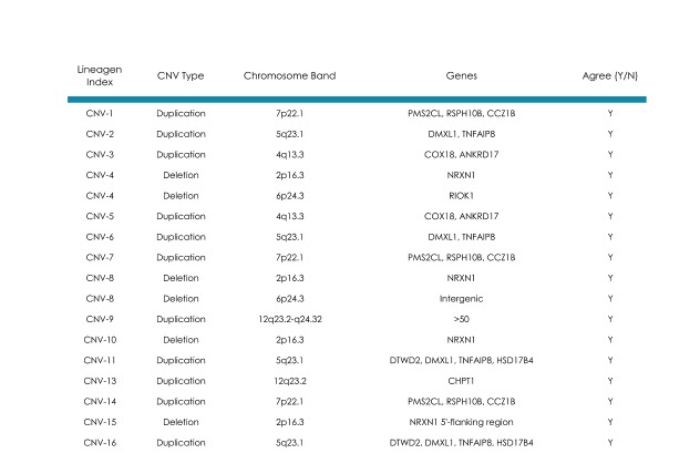
Table II: Research findings validated on FirstStepDx PLUS Samples derived from research studies with significant CNVs present15 which spanned both custom and standard probes were evaluated independently on FSDX in two laboratories and evaluated for agreement with the research findings as well their inter-laboratory concordance.
A total of six samples from patients having clinically significant CNVs findings on prior clinical analysis were re-analyzed using FSDX. This study mirrors ACMG guidance on clinical validation of a new version of a previously established platform when the total probe content change is less than 5% (here 3.3% increase)28. There was complete clinical concordance between the initial clinical results and the results generated with FSDX in two independent laboratories (AGI and CUMC). Although the cross-platform and cross-laboratory results are unequivocally clinically concordant, minor differences in breakpoint determinations were observed in the majority of analyses (8 of 11), which is expectable and reasonable within the limits of the technology and interpretation overall. The differences in breakpoints between FSDX and CytoScanHD resulted in CNVs that were smaller, but only on average by <0.25% of the total CNV size. It was previously observed that arrays with increasing increased probe density result in smaller estimates of total CNV size presumably through the higher resolving power with increased probe density14. In contrast, only a single inter-laboratory analysis of FSDX differed in breakpoint calls by the different cytogeneticists, and in this case the change was only 0.08% of the overall CNV size.
As further demonstration of the clinical validity of this array, 16 samples from an earlier research study using an alternative technology platform (Illumina, San Diego, CA)15 were analyzed on FSDX in two participating CLIA-laboratories: ARUP & Asuragen (Table II). Samples all had significant CNVs present, which had been validated by quantitative PCR in the research study, and spanned both custom and standard probes. All results were concordant both across platforms and between laboratories.
Inter-laboratory Clinical Performance Validation
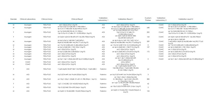
Further evidence of the inter-laboratory performance (Table III) is shown on twelve additional patients with clinically significant CNVs detected by clinical testing with FSDX at Asuragen, and then re-analyzed by both AGI and CUMC, again with completely concordant results. In addition, two patients run clinically at CUMC were concordant with results subsequently generated by AGI, and six patients run clinically at AGI were concordant with results generated by Fullerton Genetics Laboratory Center.
Table III: Inter-laboratory validation of the FirstStepDx PLUS array Independent samples from patients analyzed clinically at our participating laboratories were re-analyzed at other participating laboratories. Laboratory designations are as described in Table II.
These data clearly demonstrate the ability of FSDX to detect copy number variants on a consistent basis in independent laboratories, across a range of genomic locations with all samples yielding concordant results to those performed on three separate array platforms. The excellent performance in cases with CNVs previously detected in regions with custom probes supports the overall clinical consistency and appropriate performance of the custom probe content on the array.
Extended Comparative Clinical Sensitivity Studies in a Real-World Clinical Population
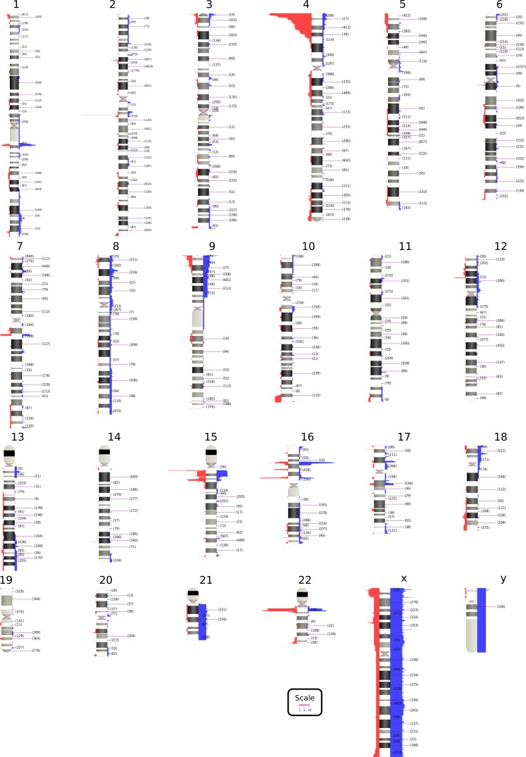
Data from 7570 consecutive patient samples tested clinically with FSDX from July 2012 (when the optimized FirstStepDx PLUS (FSDX) microarray was implemented into routine use) through May 2016 are shown in Figure 1. Overall there were 717 (9.5%) pathogenic abnormalities and 1534 (20.2%) VOUS observed, or a 29.7% overall CNV diagnostic yield for potentially abnormal findings. We also compared these results to patients who were tested by Lineagen through the same referral channels and in comparable patient cohorts and general time windows using the 2.7M and CytoScanHD arrays. The 2.7M arrays (n=378) detected 7.4% pathogenic and 8.5% VOUS for an overall yield of 15.9%. The higher density CytoScanHD arrays (n=1194) detected 9.0% pathogenic and 14.2% VOUS for an overall yield of 23.2%. It is clear that these incremental probe additions translate into potentially clinically significant yields.
Summary of clinical CNV findings using FirstStepDx PLUS.
Reported clinical findings are displayed next to chromosome Reported clinical findings are displayed next to chromosome ideograms. Deletions are shown on the left in red, and duplications are shown on the right in blue. Numbers in parentheses after chromosome band labels indicate the number of custom-designed probes in those bands. An excess of abnormalities is observed in the 4p region due to a research cohort; however this data is not reflected in the overall detection rates cited in the text.
Figure 1 visually summarizes the genomic range of CNVs detected to date using FSDX. These include known microdeletion and microduplication syndrome regions as well as variants of unknown significance.
Quality Control
Similar to the CytoScanHD, the independent analysis of CNVs with oligonucleotide and SNP probes provides both detection and confirmation simultaneously14. Three empirically determined quality control metrics are used that reflect overall data quality on an Affymetrix array: 1) waviness-SD, 2) median of the absolute values of all pairwise differences (MAPD), and 3) SNPQC (measure of how well genotype alleles are resolved). Details regarding these quality control features can be found in the Affymetrix ChAS User Guide (http://www.affymetrix.com/support/downloads/manuals/chas3_1_userguide.pdf). The criteria determined empirically for the CytoScanHD array extend to the FSDX array by virtue of design, with all three needed to meet minimum requirements for an array to be analyzed. FSDX arrays meeting these requirements can be interpreted over 99% of the time.
Analytic Validity
The results in this section are intended to demonstrate the functionality of custom probes that have been added to a microarray that is already in broad clinical use. Our goal was to show that these probes function within the parameters of standard probes on the array and respond in the expected manner to increases in input DNA.
To rigorously demonstrate the comparable analytic validity of FSDX, we employed an independent tool, the Golden Helix, Inc. Copy Number Analysis Method (CNAM) (Golden Helix, Inc. Bozeman, MT)36. First, Affymetrix Chromosome Analysis Suite (ChAS) version 2.1 (Affymetrix, Santa Clara, CA) was used to create an evaluation set of 205 samples with no reportable clinical finding from a single laboratory. To characterize the analytical function of custom probes added to the FSDX microarray, we used the sum of raw probe signal data, from these 205 samples, for all probes on across the array as a proxy for input DNA concentration. Across the 205 samples, there was roughly a 4.6-fold difference in total array signal, which we interpreted as a 4.6-fold range of input probe DNA amount. We then plotted individual probe signal intensity vs. total array signal for all probes on the array. Finally, we used regression analysis to calculate slopes and y-intercepts for each probe. To simplify the X axis, we divided total intensity by 933,696,453 (the lowest total intensity sum in the 205 samples) to generate an X-axis that ranged from 1 to 4.6. Finally, we plotted y-intercept vs. slope for each probe and compared custom probes to CytoScanHD probes. Figure 2 shows a scatter-plot based on data from this set of 205 samples. Data in red reflect custom probes only found on FSDX, while data in black designate standard CytoScanHD probes. The plot indicates that there is significant overlap between the intensity signals of the custom probes and standard probes. However, at the extreme left of the plot, some custom probes with slope near zero or negative do not appear to respond well to increasing DNA input. One can also observe in this figure that the average signal for custom probes is lower than the average signal for standard probes. These data indicate that some of the custom probes are less likely to be strongly copy number responsive, but are not necessarily non-informative. We estimate this “sub-par” population to represent approximately 20% of the custom FSDX probes. This shift in performance characteristics of the single-pass design custom probes is still within the overall desired operating parameters (note the overlap with standard probe signals in Fig. 2). Assuming that 20% of the custom probes are non-functional, custom probe content, the data suggest at most a 0.64% (% poorly functioning probes over total probe content) deviation in overall analytical sensitivity. However, given that the total probe content on FSDX is 3.27% greater, these data suggest a net gain in sensitivity of 2.63% (net increase in total optimally performing probes compared to CytoScanHD).
Functional Overlap of Custom Probes with CytoScanHD Probes.
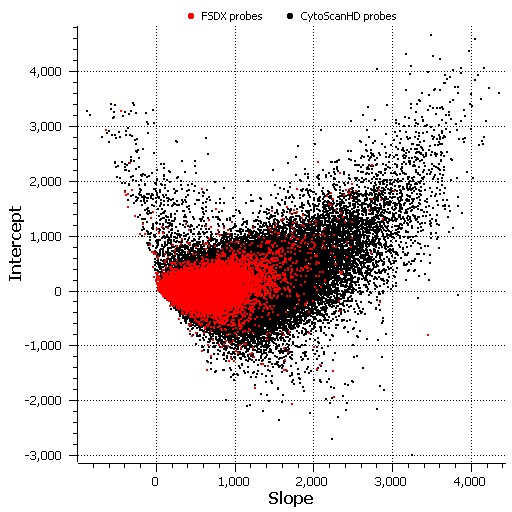
Histogram of slope and Y-intercept for each probe on the FirstStep Dx PLUS array. Probes shown in red are custom probes found only on FSDX, which overlie probes shown in black common to both FSDX and CytoScanHD. The slope for each probe indicates the change in signal relative to increases in DNA input. Probes functioning appropriately will have a positive slope. Y intercept values serve as an approximation to the background probe intensity when no sample is added to the array.
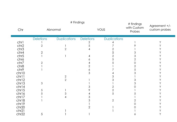
We next analyzed the relative impact of the custom probes compared to the standard array probes on overall detection rates of CNVs. The weighted log2 ratios from 184 arrays that each had at least one clinically reported finding previously analyzed using ChAS in a single laboratory were evaluated again with CNAM36. This utilized the univariate option with no moving window; a maximum of 10 segments per 10,000 probes; a minimum segment size of 1 marker; and a stringent permutation p-value threshold of 0.001. After segmentation, we classified segments as losses if their mean was <0.20 with 25 or more probes and classified segments as gains if their mean was >0.20 with 50 or more probes (consistent with our clinical workflows). These calls were compared to the clinically reported findings generated with ChAS. The analysis with CNAM was performed in two ways. First, only probes present on the CytoScanHD standard array were considered. Second, FSDX custom probes in addition to the standard probes were considered. All clinically relevant findings were observed using both analysis methods (Table IV). Each reported CNV was detectable independent of the presence of custom probes in the CNV and independent of the use of the custom probes in the analysis. These data show no evidence that the previously described sub-optimally responding subset of custom probes interfere with the function of responsive probes, whether standard or custom, or with the overall sensitivity for CNV detection.
Table IV: Analytic validity of FirstStepDxPLUS Data from clinical samples were evaluated using CNAM on weighted log2 ratios from 184 arrays as described above. The data from CNVs with and without custom FSDX probe content were evaluated to determine any discrepancy in detection based on inclusion of the custom probes and no evidence of non-concordance was observed.
The data in Table IV also demonstrate the analytic validity of custom probes that are included in some of the clinically reported findings. Increased sensitivity and resolution has been demonstrated with increasing probe density14, and since the custom probes were added specifically to regions important for ASD and other neurodevelopmental disorders, the resolution of FSDX for these conditions is predicted to be enhanced, since approximately 80% of the custom probes are fully analytically responsive to DNA input. Preliminary analysis (data not shown) suggests that the overall detection of CNVs may be increased by inclusion of the custom probes in this analysis, and further evaluation of this is under investigation.
Clinical Utility
The goal of any diagnostic test is improvement in medical management of patients and overall improvement in patient outcomes. This is achieved first by reaching the correct diagnosis and second by following the appropriate management and surveillance procedures for that diagnosis. Although there are no published reviews that analyze outcome data for patients who received a genetic diagnosis through CMA, a clear and positive impact on medical management has been documented in several studies8 , 9 , 10 , 11 , 12 , 13. Further, CMA testing often results in a correct, additional, or modified diagnosis for those who are difficult to diagnose with clinical observation alone41 , 42.
More importantly, buccal sampling in some of these patients has revealed mosaicism that would not have been detected using blood samples as a DNA source38. These findings are consistent with previous publications on mosaic FMR1 repeat expansions43, mutations in Cornelia de Lange syndrome44 and chromosomal abnormalities in Pallister-Killian syndrome45.
Even in patients with well-defined conditions, better determining CNV breakpoints with a higher resolution methodology can provide information beyond what is known or assumed from other tests46. A test which allows us to identify multiple individually rare diagnoses – such as CMA – is difficult to assess by traditional measures of clinical utility. This is due, in part, to the fact that each potential diagnosis has specific benefits in terms health and cost of care for a given patient. However, the literature has documented numerous improvements in care8 , 9 , 10 , 11 , 12 , 13 as a result of reaching a genetic diagnosis for individuals with DD, ID, and/or ASD. Further, this represents a continuum as clinical understanding and care evolves stepwise as a direct result of improved diagnostic clarity. The ability of increased probe density in genomic regions of interest to improve diagnosis is becoming apparent, and each additional correct diagnosis allows incremental opportunities for increased composite clinical utility of such tools.
Limitations
Given the rarity of individual CNVs and current limits of understanding, we report a relatively small clinical validation cohort. This could limit the certainty and range of conclusions from this evaluation; however, it exceeds established guidelines for such validations28. Analytical validity across millions of data points, as in a CMA, can only be assessed by in silico methods such as applied here. While this may be less than ideal, it is superior to mere presumptions of performance typical in the genomic literature.
CMA has proven an important clinical diagnostic advance but is limited in its ability to detect a diagnosis in a majority of affected individuals even with the increased performance and added probe content on FSDX. However, emerging evidence suggests that another significant portion of these cases (those without a diagnosis from CMA) will have mutations detectable by massively parallel sequencing (NGS) in the future47. In addition, roughly two-thirds of reported CNVs are classified as a VOUS, largely due to the limitations of our clinical experience with these rare conditions. The use of new informatics approaches and databases is beginning to better define the potential relevance and pathogenesis of such currently uncertain findings48.
Conclusions
Analytic validation of FirstStepDx PLUS coupled with over three years of clinical use have demonstrated the utility of FirstStepDx PLUS in identifying genetic causes for neurodevelopmental conditions. The simple non-invasive sampling procedure, high clinical sensitivity and extensive support services make FirstStepDx PLUS an ideal choice for the clinical genetic evaluation of patients with these disorders.
Samples and Methods
All samples were referred to Lineagen for routine clinical microarray testing. All participants, or their representatives, provided written informed consent to use the samples for research purposes (i.e., test development and validation) at the time of testing. The data collected here are covered by a protocol reviewed by the Western Institutional Review Board (WIRB protocol #20162032). No identifiers were used for any of the data evaluated in this work.
Clinical analyses were performed using Chromosome Analysis Suite v2.0.1 (Affymetrix, Inc., Santa Clara, CA). Analysis of probe function utilized Copy Number Analysis Module (Golden Helix, Inc., Bozeman, MT).
Competing Interests
The authors received funding from Lineagen, Inc. and 23&Me (commercial companies), and from Affiliated Genetics, Inc. and Columbia University Medical Center (contract service laboratories performing FSDX tests) for this study. There are no patents, products in development or marketed products to declare. This does not alter our adherence to all the PLOS ONE policies on sharing data and materials.
Corresponding Author
Charles Hensel: chensel@lineagen.com
Data Availability
Data used in this analysis are available from the NCBI dbGaP repository (https://www.ncbi.nlm.nih.gov/gap) under accession number phs001308.v1.p1.
Acknowledgments
We thank Kenny Lentz for database management and support, Porter Rickabaugh for development of the ideogram including FSDX clinical findings, and the entire Lineagen team for their remarkable energy and dedication to those with neurodevelopmental disabilities. We also thank the professional and laboratory staff of the laboratories that participated in this work.
Biographies
Director of Operations
Dr. Peiffer is a paid consultant to Lineagen and is a faculty member at the University of Utah in the Division of Medical Genetics.
Dr. Wassman is Chief Medical Officer of Lineagen, Inc., which focuses on optimized genetic diagnostic services for individuals with autism and other conditions of childhood development. A pioneer of personalized medicine, he has driven the translation and delivery of cutting-edge diagnostic technologies to clinical service for over 30-years. He has held numerous executive positions in the field of genetic testing, co-founded Good Start Genetics, and was CMO there and at Genzyme Genetics, Rosetta Genomics, Generation Health, Helicos Biosciences, Celula, and Alfigen. He is a graduate of Yale University and Albany Medical College and Board Certified in Pediatrics and Medical Genetics.
Funding Statement
The work described was funded entirely by Lineagen, Inc., the developer of FSDX. Lineagen provided support in the form of salaries for authors CH, RV, MM, SD, KH, MS, and ERW. Author AP is an employee of the University of Utah and a paid consultant to Lineagen. Authors LN and KW are employees of Affiliated Genetics, Inc., a contract service laboratory performing FSDX tests. Author BL is an employee of Columbia University Medical Center, a contract service laboratory performing FSDX tests. Author SS is a paid consultant to Lineagen, a current employee of 23&Me, and was previously an employee of ARUP, a contract service laboratory conducting tests for Lineagen at the time of this study. Author CL is a faculty member at the University of New Mexico and a paid consultant to Lineagen. These funders did not have any additional role in the study design, data collection and analysis, decision to publish, or preparation of the manuscript.
Contributor Information
Charles Hensel, Lineagen, Inc., Salt Lake City, Utah, USA.
Rena Vanzo, Clinical Genetic Services, Lineagen, Inc., Salt Lake City, Utah, USA.
Megan Martin, Lineagen, Inc., Salt Lake City, Utah, USA.
Sean Dixon, Operations, Lineagen, Inc., Salt Lake City, Utah, USA.
Christophe Lambert, Department of Internal Medicine, Center for Global Health, Division of Translational Informatics, University of New Mexico Health Sciences Center, Albuquerque, New Mexico, USA.
Brynn Levy, Department of Pathology & Cell Biology, Columbia University Medical Center, New York, New York, USA.
Lesa Nelson, Affiliated Genetics Laboratory, Inc., Salt Lake City, Utah, USA.
Andy Peiffer, Department of Pediatrics, University of Utah, Salt Lake City, Utah, USA; Lineagen, Inc., Salt Lake City, Utah, USA.
Karen S. Ho, Department of Pediatrics, University of Utah, Salt Lake City, Utah, USA; Lineagen, Inc., Salt Lake City, Utah, USA
Patricia Rushton, Lineagen, Inc., Salt Lake City, Utah, USA.
Moises Serrano, Lineagen, Inc., Salt Lake City, Utah, USA.
Sarah South, ARUP Laboratories, Salt Lake City, Utah, USA; 23andMe, Inc., Mountain View, California, USA.
Kenneth Ward, Affiliated Genetics Laboratory, Inc., Salt Lake City, Utah, USA.
Edward Wassman, Lineagen, Inc., Salt Lake City, Utah, USA.
References
- 1.Boyle CA, Boulet S, Schieve LA, Cohen RA, Blumberg SJ, Yeargin-Allsopp M, Visser S, Kogan MD. Trends in the Prevalence of Developmental Disabilities in US Children, 1997–2008. Pediatrics 2011; 127(6):1034-1042. PMID: 21606152 [DOI] [PubMed]
- 2.Manning M, Hudgins L. Array-based technology and recommendations for utilization in medical genetics practice for detection of chromosomal abnormalities. Genet Med 2010; 12(11):742-745. PMID: 20962661 [DOI] [PMC free article] [PubMed]
- 3.Schaefer GB, Mendelsohn NJ. Clinical genetics evaluation in identifying the etiology of autism spectrum disorders: 2013 guideline revisions. Genet Med 2013; 15(5):399-407. PMID: 23519317 [DOI] [PubMed]
- 4.Volkmar F, Siegel M, Woodbury-Smith M, King B, McCracken J, State M. Practice Parameter for the Assessment and Treatment of Children and Adolescents with Autism Spectrum Disorder. J Am Acad Child Adolesc Psychiatry 2014; 53(2):237-257. PMID: 24472258 [DOI] [PubMed]
- 5.Moeschler JB, Shevell M. Comprehensive Evaluation of the Child with Intellectual Disability or Global Developmental Delays. Pediatrics 2014; 134(3):e903-e918. PMID: 25157020 [DOI] [PMC free article] [PubMed]
- 6.Michelson DJ, Shevell MI, Sherr EH, Moeschler JB, Gropman AL, Ashwal S. Evidence Report: Genetic and metabolic testing on children with global developmental delay. Report of the Quality Standards Subcommittee of the American Academy of Neurology and the Practice Committee of the Child Neurology Society. Neurology 2011; 77(17):1629-1635. PMID: 21956720 [DOI] [PubMed]
- 7.Heil KM, Schaaf CP. The Genetics of Autism Spectrum Disorders – A Guide for Clinicians. Curr Psychiatry Rep 2013; 15(1):334. PMID: 23250815 [DOI] [PubMed]
- 8.Saam J, Gudgeon J, Aston E, Brothman AR. How physicians use array comparative genomic hybridization results to guide patient management in children with developmental delay. Genet Med 2008; 10(3):181-186. PMID: 18344707 [DOI] [PubMed]
- 9.Coulter ME, Miller DT, Harris DJ, Hawley P, Picker J, Roberts AE, Sobeih MM, Irons M. Chromosomal microarray testing influences medical management. Genet Med 2011; 13(9):770-776. PMID: 21716121 [DOI] [PubMed]
- 10.Ellison JW, Ravnan JB, Rosenfeld JA, Morton SA, Neill NJ, Williams MS, Lewis J, Torchia BS, Walker C, Traylor RN, Moles K, Miller E, Lantz J, Valentin C, Minier SL, Leiser K, Powell BR, Wilks TM, Shaffer LG. Clinical Utility of Chromosomal Microarray Analysis. Pediatrics 2012; 130(5):e1085-e1095. PMID: 23071206 [DOI] [PubMed]
- 11.Riggs ER, Wain KE, Riethmaier D, Smith-Packard B, Faucett WA, Hoppman N, Thorland EC, Patel VC, Miller DT. Chromosomal Microarray Impacts Clinical Management. Clin Genet 2013; 85(2):147-153. PMID: 23347240 [DOI] [PubMed]
- 12.Henderson LB, Applegate CD, Wohler E, Sheridan MB, Hoover-Fong J, Batista DA. The impact of chromosomal microarray on clinical management: a retrospective analysis. Genet Med 2014; 16(9):657-664. PMID: 24625444 [DOI] [PubMed]
- 13.Tao VQ, Chan KY, Chu YW, Mok GT, Tan TY, Yang W, Lee SL, Tang WF, Tso WW, Lau ET, Kan AS, Tang MH, Lau YL, Chung BH. The Clinical Impact of Chromosomal Microarray on Paediatric Care in Hong Kong. PLoS ONE 2014; 9(10):e109629. PMID: 25333781 [DOI] [PMC free article] [PubMed]
- 14.Mason-Suares H, Kim W, Grimmett L, Williams ES, Horner VL, Kunig D, Goldlust IS, Wu BL, Shen Y, Miller DT, Martin CL, Rudd MK. Density matters: comparison of array platforms for detection of copy-number variation and copy-neutral abnormalities. 2013; 15(9):706-712. PMID: 23558256 [DOI] [PubMed]
- 15.Matsunami N, Hadley D, Hensel CH, Christensen GB, Kim C, Frackelton E, Thomas K, da Silva RP, Stevens J, Baird L, Otterud B, Ho K, Varvil T, Leppert T, Lambert CG, Leppert M, Hakonarson H. Identification of rare recurrent copy number variants in high-risk autism families and their prevalence in a large ASD population. PLoS One 2013; 8(1):e52239. PMID: 23341896 [DOI] [PMC free article] [PubMed]
- 16.Sebat J, Lakshmi B, Malhotra D, Troge J, Lese-Martin C, Walsh T, Yamrom B, Yoon S, Krasnitz A, Kendall J, Leotta A, Pai D, Zhang R, Lee YH, Hicks J, Spence SJ, Lee AT, Puura K, Lehtimäki T, Ledbetter D, Gregersen PK, Bregman J, Sutcliffe JS, Jobanputra V, Chung W, Warburton D, King MC, Skuse D, Geschwind DH, Gilliam TC, Ye K, Wigler M. Strong Association of De Novo Copy Number Mutations with Autism. Science 2007; 316(5823):445–449. PMID: 17363630 [DOI] [PMC free article] [PubMed]
- 17.Marshall CR, Noor A, Vincent JB, Lionel AC, Feuk L, Skaug J, Shago M, Moessner R, Pinto D, Ren Y, Thiruvahindrapduram B, Fiebig A, Schreiber S, Friedman J, Ketelaars CE, Vos YJ, Ficicioglu C, Kirkpatrick S, Nicolson R, Sloman L, Summers A, Gibbons CA, Teebi A, Chitayat D, Weksberg R, Thompson A, Vardy C, Crosbie V, Luscombe S, Baatjes R, Zwaigenbaum L, Roberts W, Fernandez B, Szatmari P, Scherer SW. Structural Variation of Chromosomes in Autism Spectrum Disorder. Am J Hum Genet 2008; 82(2):477–488. PMID: 18252227 [DOI] [PMC free article] [PubMed]
- 18.Christian SL, Brune CW, Sudi J, Kumar RA, Liu S, Karamohamed S, Badner JA, Matsui S, Conroy J, McQuaid D, Gergel J, Hatchwell E, Gilliam TC, Gershon ES, Nowak NJ, Dobyns WB, Cook EH Jr. Novel Submicroscopic Chromosomal Abnormalities Detected in Autism Spectrum Disorder. Biol Psychiatry 2008 63(12):1111–1117. PMID: 18374305 [DOI] [PMC free article] [PubMed]
- 19.Glessner JT, Wang K, Cai G, Korvatska O, Kim CE, Wood S, Zhang H, Estes A, Brune CW, Bradfield JP, Imielinski M, Frackelton EC, Reichert J, Crawford EL, Munson J, Sleiman PM, Chiavacci R, Annaiah K, Thomas K, Hou C, Glaberson W, Flory J, Otieno F, Garris M, Soorya L, Klei L, Piven J, Meyer KJ, Anagnostou E, Sakurai T, Game RM, Rudd DS, Zurawiecki D, McDougle CJ, Davis LK, Miller J, Posey DJ, Michaels S, Kolevzon A, Silverman JM, Bernier R, Levy SE, Schultz RT, Dawson G, Owley T, McMahon WM, Wassink TH, Sweeney JA, Nurnberger JI, Coon H, Sutcliffe JS, Minshew NJ, Grant SF, Bucan M, Cook EH, Buxbaum JD, Devlin B, Schellenberg GD, Hakonarson H. Autism genome-wide copy number variation reveals ubiquitin and neuronal genes. Nature 2009 459(7246):569–573. PMID: 19404257 [DOI] [PMC free article] [PubMed]
- 20.Bucan M, Abrahams BS, Wang K, Glessner JT, Herman EI, Sonnenblick LI, Alvarez Retuerto AI, Imielinski M, Hadley D, Bradfield JP, Kim C, Gidaya NB, Lindquist I, Hutman T, Sigman M, Kustanovich V, Lajonchere CM, Singleton A, Kim J, Wassink TH, McMahon WM, Owley T, Sweeney JA, Coon H, Nurnberger JI, Li M, Cantor RM, Minshew NJ, Sutcliffe JS, Cook EH, Dawson G, Buxbaum JD, Grant SF, Schellenberg GD, Geschwind DH, Hakonarson H. Genome-Wide Analyses of Exonic Copy Number Variants in a Family-Based Study Point to Novel Autism Susceptibility Genes. PLoS Genet 2009 5(6):e1000536. PMID: 19557195 [DOI] [PMC free article] [PubMed]
- 21.Pinto D, Pagnamenta AT, Klei L, Anney R, Merico D, Regan R, Conroy J, Magalhaes TR, Correia C, Abrahams BS, Almeida J, Bacchelli E, Bader GD, Bailey AJ, Baird G, Battaglia A, Berney T, Bolshakova N, Bölte S, Bolton PF, Bourgeron T, Brennan S, Brian J, Bryson SE, Carson AR, Casallo G, Casey J, Chung BH, Cochrane L, Corsello C, Crawford EL, Crossett A, Cytrynbaum C, Dawson G, de Jonge M, Delorme R, Drmic I, Duketis E, Duque F, Estes A, Farrar P, Fernandez BA, Folstein SE, Fombonne E, Freitag CM, Gilbert J, Gillberg C, Glessner JT, Goldberg J, Green A, Green J, Guter SJ, Hakonarson H, Heron EA, Hill M, Holt R, Howe JL, Hughes G, Hus V, Igliozzi R, Kim C, Klauck SM, Kolevzon A, Korvatska O, Kustanovich V, Lajonchere CM, Lamb JA, Laskawiec M, Leboyer M, Le Couteur A, Leventhal BL, Lionel AC, Liu XQ, Lord C, Lotspeich L, Lund SC, Maestrini E, Mahoney W, Mantoulan C, Marshall CR, McConachie H, McDougle CJ, McGrath J, McMahon WM, Merikangas A, Migita O, Minshew NJ, Mirza GK, Munson J, Nelson SF, Noakes C, Noor A, Nygren G, Oliveira G, Papanikolaou K, Parr JR, Parrini B, Paton T, Pickles A, Pilorge M, Piven J, Ponting CP, Posey DJ, Poustka A, Poustka F, Prasad A, Ragoussis J, Renshaw K, Rickaby J, Roberts W, Roeder K, Roge B, Rutter ML, Bierut LJ, Rice JP, Salt J, Sansom K, Sato D, Segurado R, Sequeira AF, Senman L, Shah N, Sheffield VC, Soorya L, Sousa I, Stein O, Sykes N, Stoppioni V, Strawbridge C, Tancredi R, Tansey K, Thiruvahindrapduram B, Thompson AP, Thomson S, Tryfon A, Tsiantis J, Van Engeland H, Vincent JB, Volkmar F, Wallace S, Wang K, Wang Z, Wassink TH, Webber C, Weksberg R, Wing K, Wittemeyer K, Wood S, Wu J, Yaspan BL, Zurawiecki D, Zwaigenbaum L, Buxbaum JD, Cantor RM, Cook EH, Coon H, Cuccaro ML, Devlin B, Ennis S, Gallagher L, Geschwind DH, Gill M, Haines JL, Hallmayer J, Miller J, Monaco AP, Nurnberger JI Jr, Paterson AD, Pericak-Vance MA, Schellenberg GD, Szatmari P, Vicente AM, Vieland VJ, Wijsman EM, Scherer SW, Sutcliffe JS, Betancur C. Functional impact of global rare copy number variation in autism spectrum disorders. Nature 2010; 466(7304):368–372. PMID: 20531469 [DOI] [PMC free article] [PubMed]
- 22.Szatmari P, Paterson AD, Zwaigenbaum L, Roberts W, Brian J, Liu XQ, Vincent JB, Skaug JL, Thompson AP, Senman L, Feuk L, Qian C, Bryson SE, Jones MB, Marshall CR, Scherer SW, Vieland VJ, Bartlett C, Mangin LV, Goedken R, Segre A, Pericak-Vance MA, Cuccaro ML, Gilbert JR, Wright HH, Abramson RK, Betancur C, Bourgeron T, Gillberg C, Leboyer M, Buxbaum JD, Davis KL, Hollander E, Silverman JM, Hallmayer J, Lotspeich L, Sutcliffe JS, Haines JL, Folstein SE, Piven J, Wassink TH, Sheffield V, Geschwind DH, Bucan M, Brown WT, Cantor RM, Constantino JN, Gilliam TC, Herbert M, Lajonchere C, Ledbetter DH, Lese-Martin C, Miller J, Nelson S, Samango-Sprouse CA, Spence S, State M, Tanzi RE, Coon H, Dawson G, Devlin B, Estes A, Flodman P, Klei L, McMahon WM, Minshew N, Munson J, Korvatska E, Rodier PM, Schellenberg GD, Smith M, Spence MA, Stodgell C, Tepper PG, Wijsman EM, Yu CE, Rogé B, Mantoulan C, Wittemeyer K, Poustka A, Felder B, Klauck SM, Schuster C, Poustka F, Bölte S, Feineis-Matthews S, Herbrecht E, Schmötzer G, Tsiantis J, Papanikolaou K, Maestrini E, Bacchelli E, Blasi F, Carone S, Toma C, Van Engeland H, de Jonge M, Kemner C, Koop F, Langemeijer M, Hijmans C, Staal WG, Baird G, Bolton PF, Rutter ML, Weisblatt E, Green J, Aldred C, Wilkinson JA, Pickles A, Le Couteur A, Berney T, McConachie H, Bailey AJ, Francis K, Honeyman G, Hutchinson A, Parr JR, Wallace S, Monaco AP, Barnby G, Kobayashi K, Lamb JA, Sousa I, Sykes N, Cook EH, Guter SJ, Leventhal BL, Salt J, Lord C, Corsello C, Hus V, Weeks DE, Volkmar F, Tauber M, Fombonne E, Shih A, Meyer KJ. Mapping autism risk loci using genetic linkage and chromosomal rearrangements. Nat Genet 2007; 39(3):319–328. PMID: 17322880 [DOI] [PMC free article] [PubMed]
- 23.Weiss LA, Shen Y, Korn JM, Arking DE, Miller DT, Fossdal R, Saemundsen E, Stefansson H, Ferreira MA, Green T, Platt OS, Ruderfer DM, Walsh CA, Altshuler D, Chakravarti A, Tanzi RE, Stefansson K, Santangelo SL, Gusella JF, Sklar P, Wu BL, Daly MJ. Association between Microdeletion and Microduplication at 16p11.2 and Autism. N Engl J Med 2008; 358(7):667–675. PMID: 18184952 [DOI] [PubMed]
- 24.Jacquemont ML, Sanlaville D, Redon R, Raoul O, Cormier-Daire V, Lyonnet S, Amiel J, Le Merrer M, Heron D, de Blois MC, Prieur M, Vekemans M, Carter NP, Munnich A, Colleaux L, Philippe A. Array-based comparative genomic hybridisation identifies high frequency of cryptic chromosomal rearrangements in patients with syndromic autism spectrum disorders. J Med Genet 2006; 43(11):843–849. PMID: 16840569 [DOI] [PMC free article] [PubMed]
- 25.Kearney HM, South ST, Wolff DJ, Lamb A, Hamosh A, Rao KW. American College of Medical Genetics recommendations for the design and performance expectations for clinical genomic copy number microarrays intended for use in the postnatal setting for detection of constitutional abnormalities. Genet Med 2011; 13(7):676-679. PMID: 21681105 [DOI] [PubMed]
- 26.Cerebral creatine deficiency syndrome 2
- 27.Cerebral creatine deficiency syndrome 3
- 28.South ST, Lee C, Lamb AN, Higgins AW, Kearney HM. ACMG Standards and Guidelines for constitutional cytogenomic microarray analysis, including postnatal and prenatal applications: revision 2013. Genetics in Medicine 15(11):901–909 (2013). PMID: 24071793 [DOI] [PubMed]
- 29.Jez S, Martin M, South S, Vanzo R, Rothwell E. Variants of unknown significance on chromosomal microarray analysis: parental perspectives. J Community Genet 2015; 6:343-349. PMID: 25666435 [DOI] [PMC free article] [PubMed]
- 30.Turbitt E, Halliday JL, Amor DJ, Metcalfe SA. Preferences for results from genomic microarrays: comparing parents and health care providers. Clin Genet 2015; 87:21-29. PMID: 24773164 [DOI] [PubMed]
- 31.Reiff M, Bernhardt BA, Mulchandani S, Soucier D, Cornell D, Pyeritz RE, Spinner NB. “What does it mean?”: Uncertainties in understanding results of chromosomal microarray testing. Genet Med 2012; 14(2):250-258. PMID: 22241091 [DOI] [PMC free article] [PubMed]
- 32.MacDonald JR, Ziman R, Yuen RK, Feuk L, Scherer SW. The Database of Genomic Variants: a curated collection of structural variation in the human genome. Nucleic Acids Res 2014; 42(Database issue):D986-992. PMID: 24174537 [DOI] [PMC free article] [PubMed]
- 33.33. Committee on Bioethics, Committee on Genetics, and, The American College of Medical Genetics and, Genomics Social, Ethical, and Legal Issues Committee: Ethical and Policy Issues in Genetic Testing and Screening of Children. Pediatrics Mar 2013, 131 (3) 620-622; DOI: 10.1542/peds.2012-3680. PMID: 23428972 [DOI] [PubMed]
- 34.Bernardini L, Alesi V, Loddo S, Novelli A, Bottillo I, Battaglia A, Digilio MC, Zampino G, Ertel A, Fortina P, Surrey S, Dallapiccola B. High-resolution SNP arrays in mental retardation diagnostics: how much do we gain? Eur J Human Genet 2010; 18(2):178-195. PMID: 19809473 [DOI] [PMC free article] [PubMed]
- 35.McGrew SG, Peters BR, Crittendon JA, Veenstra-Vanderweele J. Diagnostic Yield of Chromosomal Microarray Analysis in an Autism Primary Care Practice: Which Guidelines to Implement? J Autism Dev Disord 2012; 42(8):1582-1591. PMID: 22089167 [DOI] [PubMed]
- 36.Howell KB, Kornberg AJ, Harvey AS, Ryan MM, Mackay MT, Freeman JL, Rodriguez Casero MV, Collins KJ, Hayman M, Mohamed A, Ware TL, Clark D, Bruno DL, Burgess T, Slater H, McGillivray G, Leventer RJ. High resolution chromosomal microarray in undiagnosed neurological disorders. J Paediatr Child Health 2013; 49(9):716-724. PMID: 23731025 [DOI] [PubMed]
- 37.Battaglia A, Doccini V, Bernardini L, Novelli A, Loddo S, Capalbo A, Filippi T, Carey JC. Confirmation of chromosomal microarray as a first-tier clinical diagnostic test for individuals with developmental delay, intellectual disability, autism spectrum disorders and dysmorphic features. Eur J Paediatr Neurol 2013; 17(6):589-599. PMID: 23711909 [DOI] [PubMed]
- 38.Miller DT, Adam MP, Aradhya S, Biesecker LG, Brothman AR, Carter NP, Church DM, Crolla JA, Eichler EE, Epstein CJ, Faucett WA, Feuk L, Friedman JM, Hamosh A, Jackson L, Kaminsky EB, Kok K, Krantz ID, Kuhn RM, Lee C, Ostell JM, Rosenberg C, Scherer SW, Spinner NB, Stavropoulos DJ, Tepperberg JH, Thorland EC, Vermeesch JR, Waggoner DJ, Watson MS, Martin CL, Ledbetter DH. Consensus Statement: Chromosomal Microarray Is a First-Tier Clinical Diagnostic Test for Individuals with Developmental Disabilities or Congenital Anomalies. Am J Hum Genet 2010; 86(5):749-764. PMID: 20466091 [DOI] [PMC free article] [PubMed]
- 39.Shen Y, Dies KA, Holm IA, Bridgemohan C, Sobeih MM, Caronna EB, Miller KJ, Frazier JA, Silverstein I, Picker J, Weissman L, Raffalli P, Jeste S, Demmer LA, Peters HK, Brewster SJ, Kowalczyk SJ, Rosen-Sheidley B, McGowan C, Duda AW 3rd, Lincoln SA, Lowe KR, Schonwald A, Robbins M, Hisama F, Wolff R, Becker R, Nasir R, Urion DK, Milunsky JM, Rappaport L, Gusella JF, Walsh CA, Wu BL, Miller DT. Clinical Genetic Testing for Patients with Autism Spectrum Disorders. 2010; Pediatr 2010; 125(4):e727-e735. PMID: 20231187 [DOI] [PMC free article] [PubMed]
- 40.SNP & Variation Suite ™ (Version 8.x) [Software]. Bozeman, MT: Golden Helix, Inc.
- 41.Sdano MR, Vanzo RJ, Martin MM, Baldwin EE, South ST, Rope AF, Allen WP, Kearney H. Clinical Utility of Chromosomal Microarray Analysis of DNA from Buccal Cells: Detection of Mosaicism in Three Patients. J Genet Counsel 2014; 23(6):922-927. PMID: 26436922 [DOI] [PubMed]
- 42.Martin MM, Vanzo RJ, Sdano MR, Baxter AL, South ST. Mosaic Deletion of 20pter Due to Rescue by Somatic Recombination. Am J Med Genet A. 2016 Jan;170A(1):243-8. doi: 10.1002/ajmg.a.37407. PMID: 26436922 [DOI] [PubMed]
- 43.Maddalena A, Yadvish KN, Spence WC, Howard-Peebles PN. A Fragile X Mosaic Male With a Cryptic Full Mutation Detected in Epithelium But Not in Blood. Am J Med Genet 64:309-312. PMID: 8844071 [DOI] [PubMed]
- 44.Huisman SA, Redeker EJ, Maas SM, Mannens MM, Hennekam RC. High rate of mosaicism in individuals with Cornelia de Lange syndrome. J Med Genet 2013; 50(5):339-344. doi: 10.1136/jmedgenet-2012-101477. PMID: 23505322 [DOI] [PubMed]
- 45.Jamuar S, Lai A, Unger S, Nishimura G. Clinical and radiological findings in Pallister-Killian syndrome. Eur J Med Genet. 2012; 55(3):167-172. doi: 10.1016/j.ejmg.2012.01.019. PMID: 22387057 [DOI] [PubMed]
- 46.Ho KS, South ST, Lortz A, Hensel CH, Sdano MR, Vanzo RJ, Martin MM, Peiffer A, Lambert CG, Calhoun A, Carey JC, Battaglia A. Chromosomal microarray testing identifies a 4p terminal region associated with seizures in Wolf-Hirschhorn syndrome. J Med Genet 2016;0:1–8. doi:10.1136/jmedgenet-2015-103626. PMID: 26747863 [DOI] [PMC free article] [PubMed]
- 47.Jiang YH, Yuen RK, Jin X, Wang M, Chen N, Wu X, Ju J, Mei J, Shi Y, He M, Wang G, Liang J, Wang Z, Cao D, Carter MT, Chrysler C, Drmic IE, Howe JL, Lau L, Marshall CR, Merico D, Nalpathamkalam T, Thiruvahindrapuram B, Thompson A, Uddin M, Walker S, Luo J, Anagnostou E, Zwaigenbaum L, Ring RH, Wang J, Lajonchere C, Wang J, Shih A, Szatmari P, Yang H, Dawson G, Li Y, Scherer SW. Detection of Clinically Relevant Genetic Variants in Autism Spectrum Disorder by Whole Genome Sequencing, The American Journal of Human Genetics (2013), http://dx.doi.org/10.1016/j.ajhg.2013.06.012. PMID: 23849776 [DOI] [PMC free article] [PubMed]
- 48.Uddin M, Tammimies K, Pellecchia G, Alipanahi B, Hu P, Wang Z, Pinto D, Lau L, Nalpathamkalam T, Marshall CR, Blencowe BJ, Frey BJ, Merico D, Yuen RK, Scherer SW. Brain-expressed exons under purifying selection are enriched for de novo mutations in autism spectrum disorder. Nat. Genetics. Vol 46(7):742-49; 2014. PMID: 24859339 [DOI] [PubMed]
Associated Data
This section collects any data citations, data availability statements, or supplementary materials included in this article.
Data Availability Statement
Data used in this analysis are available from the NCBI dbGaP repository (https://www.ncbi.nlm.nih.gov/gap) under accession number phs001308.v1.p1.


