Abstract
Negative-pressure wound therapy (NPWT) has become the standard of care for wound management. Application of NPWT to the hand and foot is technically challenging due to difficulty in obtaining a proper air seal. The cost of the treatment is another negative factor affecting NPWT in developing countries. We describe an easy and economical technique for the application of NPWT to the extremities using collage powder and sterile surgical glove. Collagen creates a physiological interface between the wound surface and the environment and is also non-immunogenic, non-pyrogenic, hypoallergenic, and pain-free. We used this method in 20 patients. In all cases, we could achieve a proper air seal and significantly reduce the cost. The median duration of NPWT application was 9 days. This combination turned out to be a good alternative to traditional VAC with respect to the duration and cost of treatment and also ergonomically better in the management of extremity wounds.
Keywords: Negative-pressure wound therapy, Collagen, Sterile gloves, Extremity VAC
Introduction
Negative-pressure wound therapy (NPWT) has become the standard of care for the management of raw areas. In spite of the advantages, limited resources and increased cost of NPWT are the adverse factors affecting its widespread use in developing countries. Moreover, application of NPWT to the extremities has the difficulty of obtaining a proper air seal due to the peculiar morphology of the hand and foot. Ng et al. described a technique using a copolymer glove to ensure a better airtight seal for a dorsal hand wound [1]. Anthony Foo modified it using an additional glove and mesenterization of the tubing for the management of multiple finger wounds. Hemmanur and Siddha used a surgical glove and Opsite for the application of NPWT in lower and upper limb wounds [2]. Here, we are discussing an easy and economical modification of NPWT to the extremities using sterile surgical gloves and collagen powder.
Method
After debridement, complete hemostasis is achieved and wound is cleaned with saline. The surrounding area of the wound is mop dried and cleaned with surgical spirit. Collagen powder (Coltyp 1 MM, Villi and Puri Scientific, India) is applied over the raw area, and paraffin gauze is placed over it. The suction tube is made of the draining tube of a sterile urobag or Ryles tube onto which multiple holes are made. A sterilized sponge of 4 cm in thickness is tailored according to the size of the wound. A longitudinal slit is made through half the thickness of the sponge by a scalpel through which the suction tube is inserted. This is placed over the paraffin gauze, and a sterile glove of no. 8 size is stretched over it. The suction tube is taken out through the edge of the glove. Tincture benzoin is taken onto gauze and wiped over the surrounding area and the edge of the wound.
Once tincture got dried, an air seal is made. A piece of transparent adhesive tape (fix-o-mull transparent, BSN medical) folded on itself with adhesive surface outside is plastered over the adjoining skin, over which the suction tube is placed and then a large piece of transparent adhesive tape is used to cover the whole edge of the gloves and skin (Fig. 1). The suction tube is further plastered to the skin by some elastic adhesive bandage. The tube is then connected to the suction machine to check for any leaks. Shrinkage of sponge under the gloves demonstrates adequate suction of the NPWT (Fig. 2). Intermittent vacuum at −125 mmHg for 15 min is applied to the tube every 0.5 h in the ward for 3 days. A 12- to 24-h gap between changing the vacuum dressings has been observed to decrease maceration. The NPWT application was continued until a desired level of granulation tissue was formed.
Fig. 1.
Schematic diagram of the NPWT method and air seal
Fig. 2.
Shrinkage of sponge under the gloves on application of suction. a Lower extremity. b Upper extremity
We used this method for the management of extremity raw area in 20 patients between January 2015 and January 2016 at the Department of Plastic Surgery, Government Medical College, Kozhikode. The indication was trauma in 12 patients and diabetic foot wound in 8 cases. The age group ranged from 21 to 68 years. There were 13 females and 7 males. It was used for lower extremity wound in 16 cases and the rest for hand raw areas. The wound size ranged between 20 and 168 cm2. The mean wound size was 54 cm2.
Results
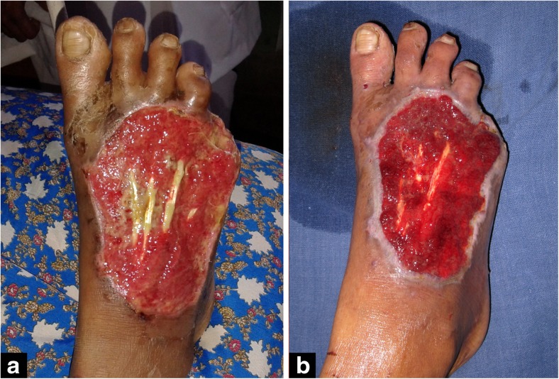
In all cases, we could achieve a proper air seal with this method. The cost could be significantly reduced to Rs.500 per NPWT. The median duration of NPWT application was 9 days. The mean total and partial dressing changes were 2.8 times. The number of NPWT applications ranged from 1 to 5. In one case, the therapy was discontinued due to patient reasons. In all other cases, wounds became clean, size was decreased, and good granulation growth could be achieved (Figs. 3 and 4).
Fig. 3.
a Raw area before NPWT. b Raw area after NPWT
Fig. 4.
a Raw area before NPWT. b Raw area after NPWT
Discussion
NPWT has become the time-tested method of wound management. This method was first described by Fleischmann et al. in 1993. Compared with traditional wound care, NPWT is believed to work based on its ability to remove edema and exudates, reduce bacteria counts, increase granulation tissue and angiogenesis, and reverse tissue retraction at an open wound. The rate of granulation tissue formation and patient compliance was better with topical negative-pressure dressing as compared to the conventional dressing [3].
The use of sterile surgical glove decreased the cost of NPWT and made it technically simple especially to attain an air seal. In extremities, it is difficult to obtain a proper air seal using an adhesive sheet. Using the glove makes this very simple since an air seal has to be created at only one end. But, the use of glove has a disadvantage of skin maceration for which NPWT is changed after every 3 days and a 12- to 24-h gap is observed between changing vacuum dressings. We used an autoclaved sponge of 4 cm in thickness for NPWT. Multiple holes were made to the sterile urobag tube to prevent blockade of the tube. Tincture benzoin application will remove the glove powder and also enhance the adhesiveness of the Opsite or fix-o-mull transparent used for the sealing of the VAC. The air seal made by the above technique was effective and remained closed for 3 days even with movements of the tube. The total cost of a single sitting of NPWT by this method was below Rs.500.
The basic principle behind NPWT is the application of sub-atmospheric pressures ranging from −25 to −200 mmHg at the wound bed. This, in turn, takes care of the factors which delay the wound care like peripheral edema and circulatory compromise at the wound bed, bacterial colonization, and retarded granulation tissue formation. NPWT creates a moist environment, reduces edema, increases local blood flow, stimulates angiogenesis and formation of granulation tissue, stimulates cell proliferation, reduces the size and complexity of the wound, removes soluble healing inhibitors from the wound, and reduces bacterial load.
Collagen is a biomaterial that encourages wound healing through deposition and organization of freshly formed fibers and granulation tissue in the wound bed, thus creating a good environment for wound healing [4]. Collagen also inhibits metalloprotienases, promotes angiogenesis, and also enhances the repair mechanisms of the body. Dr. Bhora and colleagues found that in promoting wound healing, growth factors present in collagen had certain important part. Noda et al. discovered that TGF A and TGF B present in bovine collagen were helpful in the embryonic development, cell proliferation, and tissue repair like cellular activities [5]. Collagen creates the most physiological interface between the wound surface and the environment and is impermeable to bacteria. Collagen dressing is also non-immunogenic, hemostatic, non-pyrogenic, hypoallergenic, and pain-free.
So by adding collagen powder to NPWT, the wound healing property was enhanced, thereby decreasing the duration of therapy. Patients also experienced better pain relief and compliance with this method. The use of surgical glove made the technique of acquiring an air seal simple, and also, the cost of therapy is decreased. This method is fast and ergonomic for the management of extremity wounds. Hemmanur and Siddha described the method of using surgical gloves for NPWT in 2013 [2]. We have modified this by the addition of collagen powder. To the best of our knowledge, such a combination of NPWT and collagen is not yet reported in the literature. The wound should be thoroughly debrided so that it is devoid of necrotic or infected tissue before the application of collagen powder. Further randomized control trials are required to establish the advantage of this method over the conventional method.
Conclusion
An easy and cheap alternative method of NPWT to the extremity wounds using sterile surgical gloves and collagen powder is discussed. This combination turned out to be a good alternative with respect to the duration and cost of treatment and ergonomically better in the management of extremity wounds.
Compliance with Ethical Standards
Conflict of Interest
The authors declare that they have no conflict of interest.
References
- 1.Ng R, Sebastin SJ, Tihonovs A, et al. Hand in glove—VAC dressing with active mobilisation. J Plast Reconstr Aesthet Surg. 2006;59:1011–1013. doi: 10.1016/j.bjps.2006.02.002. [DOI] [PubMed] [Google Scholar]
- 2.Hemmanur SR, Siddha LV. Role of the surgical glove in modified vacuum-assisted wound healing. Arch Plast Surg. 2013;40:630–632. doi: 10.5999/aps.2013.40.5.630. [DOI] [PMC free article] [PubMed] [Google Scholar]
- 3.Tauro LF, Ravikrishnan J, Satish Rao BS, Shenoy HD, Shetty SR, Menezes LT. A comparative study of the efficacy of topical negative pressure moist dressings and conventional moist dressings in chronic wounds. Indian J Plast Surg. 2007;40:133–140. doi: 10.4103/0970-0358.33429. [DOI] [Google Scholar]
- 4.Nataraj C, Ritter G, Dumas S, Helfer FD, Brunelle J, Sander TW. Extracellular wound matrices: novel stabilization and sterilization method for collagen-based biologic wound dressings. Wounds. 2007;19:148–156. [PubMed] [Google Scholar]
- 5.Noda, et al. Transforming growth factors A and B (TGF A & B) in bovine colostrum were involved in normal cellular activities. Gann. 1984;75:109–112. [Google Scholar]


