



Summary
Background
Primary Epstein-Barr virus (EBV) infection is usually asymptomatic, although at times it results in the benign lymphoproliferative disease, infectious mononucleosis (IM), during which almost half of patients develop hepatitis. The aims of the present study are to evaluate polymorphisms of EBV genes circulating in IM isolates from this geographic region and to investigate the correlation of viral sequence patterns with the available IM biochemical parameters.
Methods
The study included plasma samples from 128 IM patients. The genes EBNA2, LMP1, and EBNA1 were amplified using nested-PCR. EBNA2 genotyping was performed by visualization of PCR products using gel electrophoresis. Investigation of LMP1 and EBNA1 included sequence, phylogenetic, and statistical analyses.
Results
The presence of EBV DNA in plasma samples showed correlation with patients’ necessity for hospitalization (p=0.034). The majority of EBV isolates was genotype 1. LMP1 variability showed 4 known variants, and two new deletions (27-bp and 147-bp). Of the 3 analyzed attributes of LMP1 isolates, the number of 33-bp repeats less than the reference 4.5 was the only one that absolutely correlated with the elevated levels of transaminases. EBNA1 variability was presented by prototype subtypes. A particular combination of EBNA2, LMP1, and EBNA1 polymorphisms, deleted LMP1/P-thr and non-deleted LMP1/P-ala, as well as genotype 1/ 4.5 33-bp LMP1 repeats or genotype 2/ 4.5 33-bp LMP1 repeats showed correlation with elevated AST (aspartate aminotransferase) and ALT (alanine transaminase).
Conclusions
This is the first study which identified the association between EBV variability and biochemical parameters in IM patients. These results showed a possibility for the identification of hepatic related diagnostic EBV markers.
Keywords: EBV, gene polymorphism, infectious mononucleosis, LMP1, transaminase
Kratak sadržaj
Uvod
Primarna infekcija koju izaziva Epštajn-Bar virus obično je asimptomatska. Povremeno, ova infekcija može se manifestovati benignim limfoproliferativnim oboljenjem, infektivnom mononukleozom (IM), za čije vreme polovina pacijenata dobije hepatitis. Ciljevi ove studije bili su evaluacija EBV genskih polimorfizama koji su zastupljeni u IM izolatima ovog geografskog područja, kao i ispitivanje korelacije specifične virusne varijabilnosti sa dostupnim jetrinim biohemijskim parametrima IM pacijenata.
Metode
Studija je uključila uzorke plazme od 128 IM pacijenata. Za umnožavanje EBNA2, LMP1 i EBNA1 gena korišćen je nested-PCR metod. EBNA2 genotipizacija izvršena je vizuelizacijom PCR produkata korišćenjem gel elektroforeze. LMP1 i EBNA1 ispitivanje obuhvatilo je analizu njihovih genskih sekvenci, a zatim i filogenetsku i statističku obradu.
Rezultati
Prisustvo EBV DNK u uzorcima plazme korelisalo je sa neophodnošću bolničkog lečenja pacijenata (p=0,034). Većina izolata pripadala je genotipu 1. Analizom LMP1 varijabilnosti dobijene su 4 poznate varijante i 2 nove delecije (27-bp i 147-bp). Među 3 ispitivane karakteristike LMP1 izolata, broj 33-bp ponovaka manji od referenta 4,5 jedini je bio apsolutno povezan sa povišenim nivoom aspartat aminotransferaze (AST) i alanin transaminaze (ALT). EBNA1 varijabilnost dala je samo prototipne subtipove. Specifične kombinacije EBNA2, LMP1 i EBNA1 polimorfizama, LMP1 sa delecijom/P-thr i LMP1 bez delecije/P-ala, kao i genotip 1/≤4.5 33-bp LMP1 ponovka ili genotip 2/≤4.5 33-bp LMP1 ponovka, pokazale su korelaciju sa povišenim vrednostima transaminaza.
Zaključak
Ovo je prva studija u kojoj je identifikovana povezanost između EBV genetičke varijabilnosti i biohemijskih parametara IM pacijenata. Prikazani rezultati pokazuju mogućnost identifikovanja dijagnostičkih EBV markera povezanih sa statusom oštećenja jetre.
Introduction
Epstein-Barr virus (EBV) is a highly prevalent gammaherpesvirus that persistently infects more than 90% of humans by the time of adulthood. Primary EBV infection is usually asymptomatic, although at times it results in the benign lymphoproliferative disease, infectious mononucleosis (IM), especially in later childhood or young adulthood in developing countries (1). Mostly, IM is a self-limiting disease which resolves spontaneously after the emergence of EBV-specific immunity. Similar to other herpes viruses, EBV establishes a lifelong latent infection in B lymphocytes, usually without causing apparent disease in the immunocompetent host (2). However, because of its oncogenic potential, EBV can be associated with a number of malignant diseases: Burkitt’s, Hodgkin and nasal NK/T cell lymphoma, nasopharyngeal carcinoma and lymphoproliferative disorders such as posttransplantation lymphoproliferative disease (PTLD), gastric adenocarcinoma etc.
The classical presentation of IM includes fever, oropharyngitis, malaise, lymphadenopathy, hepatosplenomegaly with elevated hepatic transaminases in up to 80% of patients (3). Elevated transaminase levels are typically less than five times the upper limit of normal levels and rarely exceed 1000 U/L (4). Almost half of patients with IM have hepatitis, while jaundice is sporadic (5–10% of cases) (5). In most cases, hepatitis is benign, manifested by transient liver enzyme elevations and resolves spontaneously within 5 weeks (6). Complications like a cholestatic pattern of severe hepatitis and even fatal liver failure are rare (5).
There are no reliable parameters that predict significant liver involvement during EBV infection because the mechanism is still unclear (4). The understanding of this mechanism is complicated because it has not yet been resolved whether IM truly reflects the same events that occur in all primary EBV infections. Moreover, the majority of symptoms could be the result of other infections or even manifestations of other diseases.
Although numerous EBV-associated diseases have been described, it is unknown which EBV genome specificities contribute to the pathogenesis of these diseases. On the other hand, some geographically-associated EBV gene polymorphisms are well known and also some EBV-associated diseases are endemic to certain geographical regions. Distribution of EBV genotypes 1 and 2, mainly based on divergence within EBNA2 (EBV nuclear antigen 2) gene, shows the dominance of type 1, especially in Europe, Asia and North and South America. However, the association between genotype and disease has not been demonstrated so far (7).
Latent membrane protein 1 gene (LMP1) is an essential EBV oncogene. It transforms B cells because of the high functional similarity to the tumor necrosis factor receptor (TNFR) family members, CD40 and TNFR1 (8). The part of LMP1 gene that codes for the C-terminus of the protein shows significant variability with 7 defined LMP1 variants: Alaskan (AL), China 1, China 2, China 3, Mediterranean with (Med +) or without (Med -) deletion, and North Carolina (NC) (9, 10). In comparison with the prototype sequence (B95-8), LMP1 variants are also distinguished by presence or absence of the 30-bp deletion (30-bp-del), the number of characteristic 11 aa repeats, and defined nucleotide and amino acid (aa) changes (10). It has been suggested that LMP1 variants have different influence on the tumorigenic activity and immunogenic potential of EBV (11). Also, a geographically specific distribution of LMP1 variants has been described (12).
The only EBV gene which is expressed in all infected cells is EBNA1. Classification of EBNA1 variants includes five subtypes, two prototype sequences P-ala (B95-8 prototype) and P-thr, and three variant sequences V-val, V-leu, and V-pro. The subtype V-ala has been added afterwards (12). Subtypes are identified according to the amino acid present in locus 487 (13), and sub-variants are based on aa substitutions on loci other than the one in locus 487 (14, 15). The geographically specific distribution of EBNA1 subtypes is well-known, while reports about correlation between disease pathogenesis and EBNA1 sequence variability are debatable (14, 16).
So far, reports about an association between EBV gene polymorphisms and the clinical parameters of IM are scarce. The aims of the present study are to evaluate the polymorphisms of EBV genes circulating in IM isolates from this geographic region and to investigate the correlation of viral sequence patterns with available IM biochemical parameters.
Materials and Methods
Patients and samples
This study consists of plasma samples collected from 128 patients with infectious mononucleosis treated at the Clinic of Infectious and Tropical Diseases, Clinical Center of Serbia, collected between April 2008 and December 2010. Sample collection and research were approved by the Ethics Committee of the Faculty of Medicine, University of Belgrade, number No.29/VI-12.
Of the sample donors, 71 patients were male (55.5%), and 57 were female (44.5%). The average age was 23.5±3.2 years (15 to 38). Ninety-one (71.1%) patients had some clinical manifestations and elevation of biochemical parameters that required hospitalization: prolonged malaise, fever, sore throat, lymphadenopathy, hepatomegaly, splenomegaly, and elevated AST – aspartate aminotransferase (more than 34 U/L), ALT – alanine transaminase (more than 40 U/L) and LDH – lactate dehydrogenase (more than 333 U/L). The remaining 37 patients (28.9%) were treated in ambulatory care. Diagnosis of IM involved clinical examination, identifying atypical lymphocytes in peripheral blood smears and detection of specific anti-EBV antibodies (anti-VCA IgM, anti-VCA IgG, anti-EA-D IgG and anti-EBNA-1 IgG). Molecular investigation was performed in plasma samples of 128 patients whose results of serology screening had showed active EBV infection (primary active infection, reactivation, chronic active infection) or seronegativity, because this status did not eliminate the possibility of an active infection. Patients whose serology status showed past EBV infection did not participate in this study.
DNA isolation
Isolation of viral DNA was carried out from 200 μL plasma using a QIamp Blood Mini Kit (QIAGEN, Hilden, Germany) according to the manufacturer’s instructions. One hundred and twenty-eight DNA isolates were further used in a nested-PCR method for the amplification of three genes, EBNA2, LMP1, and EBNA1.
EBV EBNA2 genotyping
EBV genotyping was performed in 32 EBNA2 positive plasma samples, by nested-PCR as previously described (17), using primers that were reported by Mendes et al. (18). The first reaction amplified a common 596-bp region covering almost the entire EBNA2 extent, followed by two separate nested reactions amplifying distinctive regions of 497 bp for type 1 and 150 bp for type 2. EBV genotypes 1 and 2 were distinguished by identifying either the 497-bp fragment or the 150-bp fragment in gel electrophoresis (Figure 1).
Figure 1.
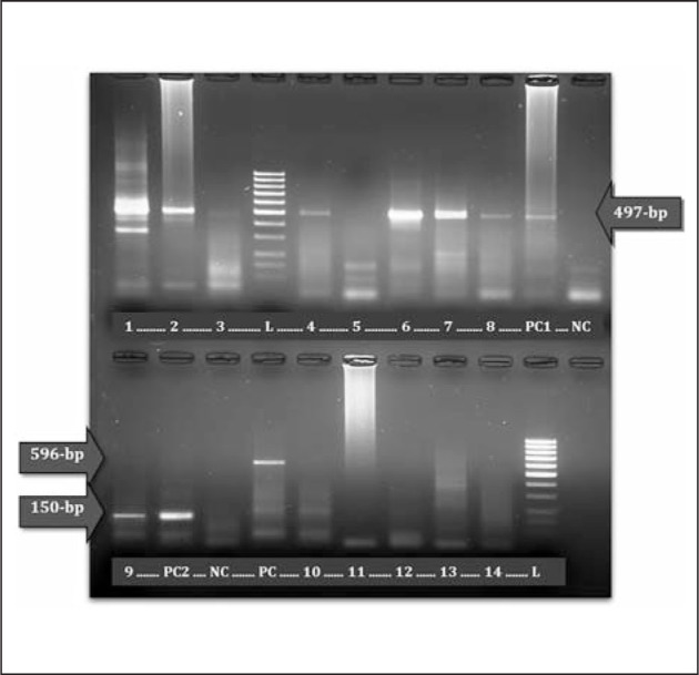
Identification of EBV genotypes 1 and 2 in gel electrophoresis: genotype 1 positive samples 1–4 and 6–8; genotype 2 positive sample – 9; negative samples 5 and 10–14; L – 100-bp standard; PC – positive control of 596-bp fragment from the first PCR reaction; PC1 – positive control of 497-bp fragment for genotype 1; PC2 – positive control of 150-bp fragment for genotype 2; NC – negative control.
LMP1 carboxy-terminal region sequencing
Amplification of the part of LMP1 gene that codes for the C terminus of the protein was performed by nested-PCR as described previously (17), using primers that were reported by Li et al. (19). Thirty-three LMP1-positive PCR products were purified using a QIAGEN MinElute Purification Kit (QIAGEN, Hilden, Germany), according to the manufacturer’s instructions. For cycle sequencing reactions, internal PCR primers and a Big Dye Terminator v 3.1 Cycle Sequencing Kit (Applied Biosystems, Foster City, CA, USA) were used. Sequencing was carried out in an automatic sequencer (ABI PRISM 310 Genetic Analyzer; Applied Biosystems, Foster City, CA, USA). Both sense and antisense strands were sequenced and compared.
EBNA1 carboxy-terminal region sequencing
Amplification of the part of EBNA1 gene that codes for the C terminus of the protein was performed by nested-PCR using primers reported by Lorenzetti et al. (15). Both PCR reactions were carried out in 40 cycles, the first reaction at: 95 °C for 1 min, 57 °C for 2 min, and 72 °C for 90 sec, and the second reaction at: 95 °C for 1 min, 60 °C for 2 min, and 72 °C for 90 sec. After the analysis of PCR products by gel electrophoresis with ethidium-bromide staining, 31 EBNA1-positive products were purified, used in cycle sequencing reactions, and sequenced based on the same principles described for LMP1 sequencing.
Sequence and phylogenetic analysis
Obtained sequences of both LMP1 and EBNA1 genes fragments (506-bp and 329-bp) were aligned and compared with a reference wild-type sequence for each gene in Bioedit 7.0.5.3 software (20). Using the same software, characteristic amino acid changes described by Edwards et al. (10) were searched for, in order to identify and classify LMP1 variants. In addition, classification of EBNA1 subtypes and sub-variants was performed after inspecting signature amino acid changes at the following positions: 471, 475, 476, 479, 487, 492, 499, 500, 502, 517, 520, 524, 525, 528 and 533.
Then, all LMP1 and EBNA1 sequences were aligned pairwise using the ClustalW method implemented in the MEGA 6.0 software (21). Corresponding reference sequences from the GenBank/EMBL/ DDBJ database were used in both alignments. The most appropriate models of evolution for the described regions of LMP1 and EBNA1 were inferred using jModelTest 2.1.4 (22). The Maximum-likelihood trees were estimated according to the defined best-fit JC+I (for EBNA1) and F81+I+G (for LMP1) evolutionary models by using the PhyML 3.0 software (23). Statistical significance of phylogeny was estimated by bootstrap analysis with 1,000 pseudoreplicate datasets. Graphical presentation and edition of phylogenetic trees were performed using Fig Tree 1.4.0 (24) and MEGA 6.0 (21) software.
Reference LMP1 and EBNA1 nucleotide sequences used in phylogenetic analysis are available in GenBank/ EMBL/DDBJ database under the accession numbers:V01555, AY493742, AY493743, AY337721, AY337722, AY493810, AY337723, AY493835, AY337724, AY493799, AY337725, AY337726, X58140, GU475455, JN986939, AF192742, GU475448, AF192743, GU475431, AF192744, JN986947 and GU475442.
Statistical analyses
The chi-squared or Fisher’s exact test and Student’s t-test were used for statistical analysis. Analyses were performed by SPSS v.21 for Windows (SPSS Inc., Chicago, IL, USA) software. P-value ≤ 0.05 was considered statistically significant.
Results
EBV DNA was detected in 33/128 plasma samples (25.8%) with similar distribution between male (28.2%) and female (22.8%) patients. Statistical analysis showed a correlation between EBV DNA presence and the patients’ necessity for hospitalization (p=0.034). Thus, EBV DNA was detected in 31.1% of hospitalized patients (28/90) and in only 13.2% of non-hospitalized patients (5/38). Transaminase (AST and ALT) levels increased in 82.8% (106/128) patients for AST and 93.7% (120/128) for ALT. There was no correlation between EBV DNA presence and elevation of biochemical parameters over normal levels: AST over 34 U/L (p=1), ALT over 40 U/L (p=0.746) or LDH over 333 U/L (p=0.360). In addition, EBV DNA presence did not correlate with hepatomegaly (p=0.065) or splenomegaly (p=0.573).
EBV genotyping
The frequency of EBV genotype 1 or genotype 2 was determined in 32 plasma samples which were positive for EBNA2 gene. Genotype 1 was present in 96.87% samples (31/32) and genotype 2 in 3.13% samples (1/32).
LMP1 sequence characterization and correlation with biochemical parameters
Thirty-three sequences of the part of EBV LMP1 gene (coordinates 168719–168213) were obtained, analyzed, and compared with the B95-8 prototype sequence. Characteristic nucleotide variability including variant characterization, detection of deletions, determination of the number of 11-amino acid repeats, and inspection of amino acid changes were investigated, followed by phylogenetic analysis.
As shown in Figure 2, the phylogenetic analysis clustered LMP1 sequences from this study along with other isolates from GenBank into 4 groups. These clusters represented 4 of the 7 known variants, namely B95-8, Med, China1 and NC. To confirm the phylogenetic grouping, it was necessary to check out variant-characteristic aa changes defined by Edwards et al. (10). The most dominant variant was China 1 (36.4%) (Table I).
Figure 2.

Phylogenetic tree of the C-terminus of LMP1. Thirty-three 506-bp fragments of LMP1 IM sequences and 13 sequences obtained from GenBank/EMBL7DDBJ database.
Table I.
Distribution of LMP1 33-bp repeat units and deletions between different LMP1 variants identified in IM isolates.
| LMP1 characteristic | LMP1 variant | Number of 33-bp tandem repeat units | LMP1 deletion | Total | ||
|---|---|---|---|---|---|---|
| 3–4.5 | 5–6 | |||||
| B95-8 | 10 | – | No del | 9 | 10 (30.3%) | |
| 27-bp-del | 1 | |||||
| China 1 | 6 | 6 | 30-bp-del | 12 | 12 (36.4%) | |
| NC | 5 | 1 | No del | 6 | 6 (18.2%) | |
| Med | – | 5 | No del | 4 | 5 (15.1%) | |
| 147-bp-del | 1 | |||||
| Total | 21 (63.6%) | 12 (36.4%) | 33 | |||
| P-value1 | <0.001 | |||||
P-value1 for significant differences in the distribution of the number of 33-bp repeats (≤4.5 or >4.5) between LMP1 variants.
LMP1 isolates were also divided into 4 groups based on the presence of specific deletions. The most frequent were non-deleted isolates (57.6%). However, three deletions were identified in almost half of all LMP1 sequences (42.4%), with the dominance of specific 10-aa/30-bp deletion (36.4%). The remaining 2 deletions, which were identified in 1 isolate each, were newly found: 9-aa/27-bp and 49-aa/147-bp (Table I).
It has been shown that the C-terminal domain of LMP1 could contain various numbers of 11-aa repeats located between aa 250 and 308 (25). The B95-8 prototype sequence has four perfect repeats with a disruption of 5-aa between the second and the third repeat (4.5 11-aa repeats). Therefore, isolates from this study were classified into two groups: those with 4.5 repeats or less, and those with more than 4.5 repeats (Table I). The number of repeats varied from 3 to 6, and the group with 4.5 repeats or less was the most common (63.6%).
The analysis of amino acid changes included identification of 7 characteristic aa positions for variant discrimination, described by Edwards et al. (10). However, 49 aa substitutions were identified at additional 39 positions and some of them were unique for NC and B95-8 variants. For NC isolates, 3 substiutions were unique: Asp Asn (position 250), Ser Pro (position 313) and Gly Gln (position 331), and for B95-8 isolates Glu Gln (position 328) substitution was unique.
When 3 LMP1 sequence attributes (variant, deletion and the presence of ≤4.5 or >4.5 33-bp repeats) were analyzed together, a significant difference was found in the distribution of the number of 33-bp repeats (≤4.5 or >4.5) between LMP1 variants (p<0.001) (Table I). Thus, B95-8 isolates always contained ≤4.5 33-bp repeats, in contrast to Med isolates which always contained >4.5 33-bp repeats. None of the 3 analyzed LMP1 sequence attributes correlated with the levels of AST, ALT, LDH or severity of clinical manifestations. However, isolates with 3 or 4 33-bp repeats were only identified in patients with elevated AST and ALT (Figure 3).
Figure 3.

Distribution of the number of 33-bp LMP1 repeats (3-6) in IM patients with different AST and ALT levels.
EBNA1 sequence characterization and correlation with biochemical parameters
Thirty-one sequences of the part of EBV EBNA1 gene (coordinates 109261-109590) were obtained, analyzed, and compared with the B95-8 prototype sequence. According to aa changes and clustering of isolates in the phylogenetic tree, 2 prototype subtypes were identified: P-ala (12/31) and P-thr (19/31) (Figure 4). Analysis of characteristic sequence variability aside from subtype-specific aa substitutions included subvariant characterization within the scope of each subtype. In contrast to all P-thr isolates which did not show any additional (subvariant) variability, all P-ala isolates belonged to P-ala subvariant 2 (P-ala-sv-2). Thus, P-ala-sv-2 had aa changes Asp Glu (on position 499) and Thr Val (on position 524), compared with the B95-8 prototype sequence (P-ala). EBNA1 sequence characteristics did not show correlations with biochemical or clinical parameters.
Figure 4.

Phylogenetic tree of the C-terminus of EBNA1. Thirty-one 329-bp fragments of EBNA1 IM sequences and 10 sequences obtained from GenBank/EMBL7DDBJ database.
Correlation between polymorphisms of EBV genes and biochemical parameters
In order to access the potential association between biochemical and clinical parameters and the comprehensive variability of 3 EBV genes, 10 different EBV polymorphisms were defined. Each polymorphism comprised a specific combination between the described characteristics of 2 or 3 EBV genes: EBNA2 genotype, LMP1 variant, the presence of LMP1 deletion, number of 33-bp LMP1 repeats (≤4.5 or >4.5) and EBNA1 subtype. The major polymorphism was determined by EBNA2 genotype, LMP1 variant and EBNA1 subtype. Among the isolates, the most frequent was genotype 1/China1/P-thr (29%). According to the accessible anamnestic and clinical data of IM patients, no correlation was found between defined EBV polymorphisms and sex, severity of clinical manifestations (hospitalized or ambulatory patients) or elevation of LDH. On the other hand, the difference in the distribution of polymorphism presence of LMP1 deletion/EBNA1 subtype between patients with normal and elevated levels of transaminases was statistically significant: AST (p=0.039) and ALT (p=0.039). Therefore, deleted LMP1/P-thr and non-deleted LMP1/P-ala were found only when both AST and ALT levels were elevated (Table II). One polymorphism more (EBNA2 genotype/number of 33-bp LMP1 repeats) showed a borderline level of statistically significant difference in presentation between patients with normal and elevated levels of AST and ALT (p=0.053). Isolates with genotype 1/ 4.5 33-bp LMP1 repeats or genotype 2/ 4.5 33-bp LMP1 repeats were mostly found in patients with elevated AST and ALT (Table II).
Table II.
Correlation between different presentations of two EBV gene polymorphisms and AST and ALT levels found in IM patients.
| Total | AST | ALT | ||||
|---|---|---|---|---|---|---|
| normal | elevated | normal | elevated | |||
| LMP1 deletion/EBNA1 subtype | Non-deleted/P-thr | 9 | 3 | 6 | 3 | 6 |
| Non-deleted/P-ala | 9 | – | 9 | – | 9 | |
| Deleted/P-thr | 10 | – | 10 | – | 10 | |
| Deleted/P-ala | 3 | 1 | 2 | 1 | 2 | |
| Total | 31 | 4 | 27 | 4 | 27 | |
| P-value1 | p=0.039 | p=0.039 | ||||
| EBNA2 genotype/number of 33-bp LMP1 repeats | Genotype 1/≤4.5 33-bp repeats | 19 | 2 | 17 | 2 | 17 |
| Genotype 1/≤4.5 33-bp repeats | 12 | 3 | 9 | 3 | 9 | |
| Genotype 2/<4.5 33-bp repeats | 1 | – | 1 | – | 1 | |
| Total | 32 | 5 | 27 | 5 | 27 | |
| P-value1 | p=0.053 | p=0.053 | ||||
P-value1 for significant or borderline level of statistically significant difference in distribution of presentations of EBV polymorphisms between patients with normal and elevated AST and ALT levels
Discussion
Hepatic involvement in infectious mononucleosis varies in severity and frequency. The incidence is estimated to be 10% in young adults and 30% in the elderly (26). Hepatotropic viruses, including EBV, are not considered to be directly cytotoxic. Instead, the immune response to viral antigens, which are expressed on hepatocytes, results in hepatocyte death. In IM, EBV infected and activated CD8+ T cells may accumulate in the liver causing hepatosplenomegaly and lymphadenopathy. The products of the EBV-infected CD8+ T cells or infiltrating cytotoxic T lymphocytes induce hepatic injury (27). Although such hepatitis is usually mild and can go undetected with spontaneous resolution, there are described cases with complications like severe hepatitis or acute hepatic failure (28). Predictors or virus specific risk factors for IM hepatic involvement remain unknown. Among patients from this study, hepatic transaminase levels increased in 82.8% of patients for AST and 93.7% for ALT, respectively. This result is similar to a number of previous reports (29).
The earliest events of IM, after initial oropharyngeal contact with EBV infected saliva, remain mostly unclear because of the late clinical presentation of IM. Detection of EBV DNA is possible in 52–100% of patients in the early stages of infection, and in less than 40% of patients in the last days of acute infection (30–32). Two to three weeks after the onset of clinical manifestations the virus is undetectable in blood (33). This onset for patients from this study was 2–28 days before visiting the hospital. Thus, in most cases, viremia was very low or undetectable in PCR testing (74.2%). Detection of EBV DNA did not correlate with sex, hepatosplenomegaly or levels of transaminases. However, it was significant that EBV DNA had been detected in more than a third (31.1%) of hospitalized patients in contrast to only 13.2% of non-hospitalized patients. It could be supposed that clinical presentations are more severe during EBV replication and detectable viremia in peripheral blood.
Coherent with worldwide genotype distribution, the majority of EBV found in Serbian isolates was genotype 1. Geographically determined distribution of EBV genotypes was earlier explained with a potential association between the genetic disposition of the human population from different geographical regions and particular EBV genotypes (7). The presence of four known LMP1 variants (B95-8, Med, China1 and NC) was in accordance with the already described European distribution based on the analysis of isolates from non-malignant and healthy patients (12). However, previously published data showed domination of B95-8 (42%) in contrast to the domination of China 1 (36%) demonstrated in this study. On the other hand, the dominance of non-deleted isolates was characteristic for the isolates of European origin (12). It is particularly interesting that IM isolates from this region displayed two newly found deletions, the 27-bp deletion and the 147-bp deletion. The deletion 147-bp has not been found earlier in Serbian isolates from malignant tissues (17). Therefore, it could be assumed that it might cover the region which was somehow involved in oncogenesis.
Recombination activity within LMP1 sequence is most notable in the scope of the 33-bp repeats site. These repeats are part of CTAR-3 region which is responsible for binding to JAK-3 and activation of JAK-3/STAT signal pathway and they do not have transformational influence on the infected cell (9). The fact that more deleted (50%) than non-deleted (35.7%) isolates had a high number of 33-bp repeats could be the consequence of intensive recombination activity during lytic replication and possibly of an unknown mechanism of nucleotide compensation after the formation of deletion (9). More important was the fact that isolates with a small number of 33-bp repeats (less than reference 4.5) were only found in patients with elevated AST and ALT levels. Although there is no evidence of functional association between the mentioned parameters, it could be assumed that the recombination within the 33-bp repeats region could affect metabolic processes within infected cells which include regulation of AST and ALT levels.
The C terminal region of LMP1, especially the part between aa 322 and 366, has been described as a mutational hot spot, because of numerous substitutions that occurred during the evolution of LMP1 variants (34). Besides 7 characteristic aa positions, 39 more positions displayed 4 unique aa changes for NC and B95-8 variants. These substitutions might serve as new markers for variant discrimination.
Besides the earlier demonstration of associations between the variability of the EBNA1 gene and geographical origin, there was also a theory about the determined tropism of specific EBNA1 subtypes (35). Identification of prototype subtypes, P-thr i P-ala, with domination of the P-thr, was similar to the previously described European EBNA1 distribution in healthy and non-malignant isolates (12), and also supported the second theory that both prototype EBNA1 sequences (P-ala or P-thr) could only be detected in healthy individuals and not in tumor tissues (13, 36). Finally, characterization of EBNA1 was completed by identification of the P-ala-sv-2 subvariant which had already been reported only in isolates from Danish non-malignant isolates (14). Therefore, this subvariant undoubtedly represents European specific EBNA1 variability (12).
Specific EBV disease characteristics might also be the result of different combinations of EBV genome variability (30). Only a few studies attempted to define the EBV polymorphisms of EBNA2, LMP1 and EBNA1 genes in specific types of disease, but no correlation was established (15, 30). Because of that, the significant associations that were found after comparative analysis of EBV genes polymorphisms and IM parameters might be of special interest. Those were correlations that combinations the presence of LMP1 deletions/EBNA1 subtype, and also EBNA2 genotype/ number of 33-bp LMP1 repeats had with levels of hepatic transaminases. Taken together, it would be essential to investigate the molecular background of progression between 4 specified EBV polymorphisms and host cell in vitro.
The characterization of EBV gene variability and identification of structural changes that might affect the functionality of gene products could explain the specific contribution of EBV variability to the pathogenesis of EBV-associated diseases. This study identified some new gene characteristics, and according to our knowledge attempted to establish an association between EBV variability and biochemical parameters in IM patients for the first time. It was shown that specific EBV polymorphisms were present only when AST and ALT levels were elevated. These results might be used for further investigation, as there is a possibility for the identification of hepatic related diagnostic EBV markers.
Acknowledgements
The study was supported by the Ministry of Education, Science and Technological Development, Republic of Serbia, Grant No. 175073. The authors are grateful to the Laboratory Technicians Gabrijela Pavlović and Marija Janković from the Virology Department, Institute of Microbiology and Immunology, Faculty of Medicine, University of Belgrade, for their technical assistance.
Glossary
List of abbreviations
- aa
amino acid
- AL
Alaskan
- ALT
alanine transaminase
- AST
aspartate aminotransferase
- EBNA
EBV nuclear antigen
- EBV
Epstein-Barr virus
- IM
infectious mononucleosis
- LDH
lactate dehydrogenase
- LMP1
latent membrane protein 1
- Med
Mediterranean
- NC
North Carolina
- sv
subvariant
- TNFR
tumor necrosis factor receptor
- del
deletion
Conflict of interest statement
The authors stated that they have no conflicts of interest regarding the publication of this article.
References
- 1.Rickinson AB, Kieff E. Knipe D, Howlry P. Fields Virology. 5th edition. Philadelphia, PA: Lippincott Williams & Wilkins; 2007. Epstein-Barr Virus and Its Replication; pp. 2603–54. [Google Scholar]
- 2.Young LS, Rickinson AB. Epstein-Barr virus: 40 years on. Nat Rev Cancer. 2004;4:757–68. doi: 10.1038/nrc1452. [DOI] [PubMed] [Google Scholar]
- 3.Markin RS. Manifestations of Epstein-Barr virus-associated disorders in liver. Liver. 1994;14:1–13. doi: 10.1111/j.1600-0676.1994.tb00001.x. [DOI] [PubMed] [Google Scholar]
- 4.Tan ZH, Phua KB, Ong C, Kader A. Prolonged hepatitis and jaundice: a rare complication of paediatric Epstein-Barr virus infection. Singapore Med J. 2015;56:e112–5. doi: 10.11622/smedj.2015113. [DOI] [PMC free article] [PubMed] [Google Scholar]
- 5.Petrova M, Kamburov V. Epstein-Barr virus: silent companion or causative agent of chronic liver disease? World J Gastroenterol. 2010;16:4130–4. doi: 10.3748/wjg.v16.i33.4130. [DOI] [PMC free article] [PubMed] [Google Scholar]
- 6.Kofteridis DP, Koulentaki M, Valachis A, Christofaki M, Mazokopakis E, Papazoglou G. et al. Epstein Barr virus hepatitis. Eur J Intern Med. 2011;22:73–6. doi: 10.1016/j.ejim.2010.07.016. [DOI] [PubMed] [Google Scholar]
- 7.Ikegaya H, Motani H, Sakurada K, Sato K, Akutsu T, Yoshino M. Forensic application of Epstein-Barr virus genotype: Correlation between viral genotype and geographical area. J Virol Methods. 2007;147:78–85. doi: 10.1016/j.jviromet.2007.08.010. [DOI] [PubMed] [Google Scholar]
- 8.Kulwichit W, Edwards RH, Davenport EM, Baskar IF, Godfrey V, Raab-Traub N. Expression of the Epstein-Barr virus latent membrane protein 1 induces B cell lymphoma in transgenic mice. Proc Natl Acad Sci USA. 1998;95:11963–8. doi: 10.1073/pnas.95.20.11963. [DOI] [PMC free article] [PubMed] [Google Scholar]
- 9.Miller WE, Edwards RH, Walling DM, Raab-Traub N. Sequence variation in the Epstein-Barr virus latent membrane protein 1. J Gen Virol. 1994;75:2729–40. doi: 10.1099/0022-1317-75-10-2729. [DOI] [PubMed] [Google Scholar]
- 10.Edwards RH, Seillier-Moiseiwitsch F, Raab-Traub N. Signature amino acid changes in latent membrane protein 1 distinguish Epstein-Barr virus strains. Virology. 1999;261:79–95. doi: 10.1006/viro.1999.9855. [DOI] [PubMed] [Google Scholar]
- 11.Knecht H, Bachmann E, Brousset P, Sandvej K, Nadal D, Bachmann F. et al. Deletions within the LMP1 oncogene of Epstein-Barr virus are clustered in Hodgkin’s disease and identical to those observed in nasopharyngeal carcinoma. Blood. 1993;82:2937–42. [PubMed] [Google Scholar]
- 12.Chang CM, Yu KJ, Mbulaiteye SM, Hildesheim A, Bhatia K. The extent of genetic diversity of Epstein-Barr virus and its geographic and disease patterns: a need for reappraisal. Virus Res. 2009;143:209–21. doi: 10.1016/j.virusres.2009.07.005. [DOI] [PMC free article] [PubMed] [Google Scholar]
- 13.Bhatia K, Raj A, Guitierrez MI, Judde JG, Spangler G, Venkatesh H. et al. Variation in the sequence of Epstein-Barr virus nuclear antigen 1 in normal peripheal blood lymphocytes and in Burkitt’s lymphomas. Oncogene. 1996;13:177–81. [PubMed] [Google Scholar]
- 14.Sandvej K, Zhou XG, Hamilton-Dutoit S. EBNA-1 sequence variation in Danish and Chinese EBV-associated tumors: evidence for geographical polymorphism but not for tumor-specific subtype restriction. J Pathol. 2000;191:127–31. doi: 10.1002/(SICI)1096-9896(200006)191:2<127::AID-PATH614>3.0.CO;2-E. [DOI] [PubMed] [Google Scholar]
- 15.Lorenzetti MA, Altcheh J, Moroni S, Moscatelli G, Chabay PA, Preciado MV. EBNA1 sequences in Argentinean pediatric acute and latent Epstein-Barr virus infection reflect circulation of novel South American variants. J Med Virol. 2010;82:1730–8. doi: 10.1002/jmv.21871. [DOI] [PubMed] [Google Scholar]
- 16.Puchhammer-Stockl E, Gorzer I. Cytomegalovirus and Epstein-Barr virus subtypes – The search for clinical significance. J Clin Virol. 2006;36:239–48. doi: 10.1016/j.jcv.2006.03.004. [DOI] [PubMed] [Google Scholar]
- 17.Banko A, Lazarevic I, Cupic M, Stevanovic G, Boricic I, Jovanovic T. Carboxy-terminal sequence variation of LMP1 gene in Epstein-Barr-virus-associated mononucleosis and tumors from Serbian patients. J Med Virol. 2012;84:632–42. doi: 10.1002/jmv.23217. [DOI] [PubMed] [Google Scholar]
- 18.Mendes TM, Oliveira LC, Yamamoto L, Del Negro GM, Okay TS. Epstein-Barr virus nuclear antigen-2 detection and typing in immunocompromised children correlated with lymphoproliferative disorder biopsy findings. Braz J Infect Dis. 2008;12:186–91. doi: 10.1590/s1413-86702008000300005. [DOI] [PubMed] [Google Scholar]
- 19.Li DJ, Bei JX, Mai SJ, Xu JF, Chen LZ, Zhang RH. et al. The dominance of China 1 in the spectrum of Epstein-Barr virus strains from Cantonese patients with nasopharyngeal carcinoma. J Med Virol. 2009;81:1253–60. doi: 10.1002/jmv.21503. [DOI] [PubMed] [Google Scholar]
- 20.Hall TA. BioEdit: A user-friendly biological sequence alignment editor and analysis program for Windows 95, 98, NT, 2000, XP. Nucl Acids Symp. 1999;41:95–8. [Google Scholar]
- 21.Tamura K, Stecher G, Peterson D, Filipski A, Kumar S. MEGA6: Molecular Evolutionary Genetics Analysis Version 6.0. Mol Biol Evol. 2013;30:2725–9. doi: 10.1093/molbev/mst197. [DOI] [PMC free article] [PubMed] [Google Scholar]
- 22.Darriba D, Taboada GL, Doallo R, Posada D. jModelTest 2: more models, new heuristics and parallel computing. Nat Methods. 2012;9:772. doi: 10.1038/nmeth.2109. [DOI] [PMC free article] [PubMed] [Google Scholar]
- 23.Guindon S, Gascuel O. A simple, fast and accurate algorithm to estimate large phylogenies by maximum likelihood. Syst Biol. 2003;52:696–704. doi: 10.1080/10635150390235520. [DOI] [PubMed] [Google Scholar]
- 24.Rambaut A. Molecular evolution, phylogenetics and epidemiology [Internet] 2007 http://tree.bio.ed.ac.uk/software/figtree/ [updated 2012 December 5; cited 2014 july 6]. Available from:
- 25.Senyuta N, Yakovleva L, Goncharova E, Scherback L, Diduk S, Smirnova K. et al. Epstein-Barr virus latent membrane protein 1 polymorphism in nasopharyngeal carcinoma and other oral cavity tumors in Russia. J Med Virol. 2014;86:290–300. doi: 10.1002/jmv.23729. [DOI] [PubMed] [Google Scholar]
- 26.Lawee D. Mild infectious mononucleosis presenting with transient mixed liver disease: case report with a literature review. Can Fam Physician. 2007;53:1314–6. [PMC free article] [PubMed] [Google Scholar]
- 27.Kang MJ, Kim TH, Shim KN, Jung SA, Cho MS, Yoo K. et al. Infectious mononucleosis hepatitis in young adults: two case reports. Korean J Intern Med. 2009;24:381–7. doi: 10.3904/kjim.2009.24.4.381. [DOI] [PMC free article] [PubMed] [Google Scholar]
- 28.Mellinger JL, Rossaro L, Naugler WE, Nadig SN, Appelman H, Lee WM. et al. Epstein-Barr virus (EBV) related acute liver failure: a case series from the US Acute Liver Failure Study Group. Dig Dis Sci. 2014;59:1630–7. doi: 10.1007/s10620-014-3029-2. [DOI] [PMC free article] [PubMed] [Google Scholar]
- 29.Yang SI, Geong JH, Kim JY. Clinical characteristics of primary Epstein Barr virus hepatitis with elevation of alkaline phosphatase and γ-glutamyltransferase in children. Yonsei Med J. 2014;55:107–12. doi: 10.3349/ymj.2014.55.1.107. [DOI] [PMC free article] [PubMed] [Google Scholar]
- 30.Ai J, Xie Z, Liu C, Huang Z, Xu J. Analysis of EBNA-1 and LMP-1 variants in diseases associated with EBV infection in Chinese children. Virol J. 2012;9:13. doi: 10.1186/1743-422X-9-13. [DOI] [PMC free article] [PubMed] [Google Scholar]
- 31.Yamamoto M, Kimura H, Hironaka T, Hirai K, Hasegawa S, Kuzushima K. et al. Detection and quantification of virus DNA in plasma of patients with Epstein-Barr virusassociated diseases. J Clin Microbiol. 1995;33:1765–8. doi: 10.1128/jcm.33.7.1765-1768.1995. [DOI] [PMC free article] [PubMed] [Google Scholar]
- 32.Cheng CC, Chang LY, Shao PL, Lee PI, Chen JM, Lu CY. et al. Clinical manifestations and quantitative analysis of virus load in Taiwanese children with Epstein-Barr virusassociated infectious mononucleosis. J Microbiol Immunol Infect. 2007;40:216–21. [PubMed] [Google Scholar]
- 33.Bauer CC, Aberle SW, Popow-Kraupp T, Kapitan M, Hofmann H, Puchhammer-Stöckl E. Serum Epstein-Barr virus DNA load in primary Epstein-Barr virus infection. J Clin Microbiol. 2005;75:54–58. doi: 10.1002/jmv.20237. [DOI] [PubMed] [Google Scholar]
- 34.Sandvej K, Peh SC, Andresen BS, Pallesen G. Identification of potential hot spots in the carboxy-terminal part of the Epstein-Barr virus (EBV) BNLF-1 gene in both malignant and benign EBV-associated diseases: high frequency of a 30-bp deletion in Malaysian and Danish peripheral T-cell lymphomas. Blood. 1994;84:4053–60. [PubMed] [Google Scholar]
- 35.Gutierrez MI, Raj A, Spangler G, Sharma A, Hussain A, Judde JG. et al. Sequence variations in EBNA-1 may dictate restriction of tissue distribution of Epstein-Barr virus in normal and tumour cells. J Gen Virol. 1997;78:1663–70. doi: 10.1099/0022-1317-78-7-1663. [DOI] [PubMed] [Google Scholar]
- 36.Gutiérrez MI, Spangler G, Kingma D, Raffeld M, Guerrero I, Misad O. et al. Epstein-Barr virus in nasal lymphomas contains multiple ongoing mutations in the EBNA-1 gene. Blood. 1998;92:600–6. [PubMed] [Google Scholar]