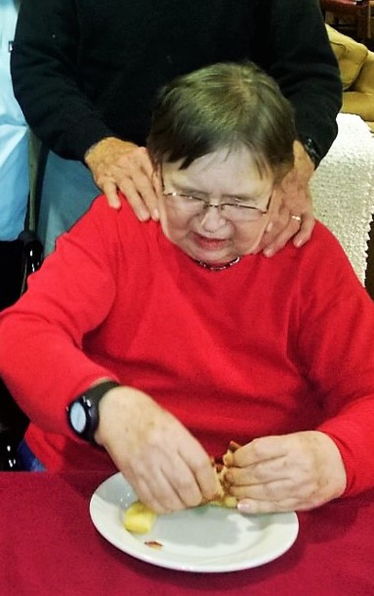
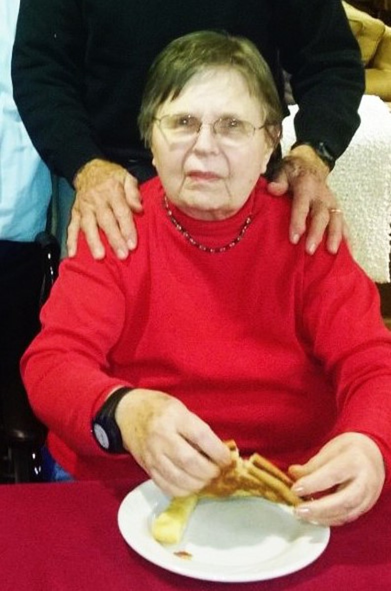
This letter updates the April 2016 case report1 about an 81-year-old patient who was in the final stages of advanced Alzheimer disease (AD) in hospice care. A neuropsychologist examined her on May 21, 2015, and concluded that she was “completely nonresponsive.” Following treatment by 4 computed tomography (CT) scans of the brain from July to August 2015, the patient made a remarkable recovery. A fifth scan on October 1 caused a setback, from which she gradually recovered. On November 20, she was judged to be no longer eligible for hospice care because her condition was sufficiently improved. Since then, she has participated in a stimulating, dementia day care program. Photos on December 4, 2015, Figures 1 and 2, demonstrate restoration of appetite and responsiveness.
Figure 1.

Restoration of appetite of patient with Alzheimer disease (AD).
Figure 2.

Response of patient with Alzheimer disease (AD) to request to look at the camera.
Recognizing that the efficacy of the CT scan treatments would likely be transitory, the patient’s spouse requested ongoing booster scans every 4 to 5 months. These started on February 24, 2016, about 21 weeks after the October 1 treatment.
Dr William D. MacInnes (note 1) reexamined the patient on April 15, 2016. His progress note states:
Unlike our last visit, Mrs XXX was able to give simple verbal responses to direct, simple questions. Not all of her responses were related to the direct questions, but she seemed to be reacting appropriately to the prosody and nonverbal cues of those around her. This represents some improvement from October 12, 2015, when I last saw her.”…“Mr XXX reported that his wife is no longer receiving services through hospice at this time because of her lack of decline. He indicated that she was able to get out of the car by herself with some standby assist. However, she has not resumed walking independently. Mr XXX reported that his wife occasionally feeds herself, but she still requires cueing.
The dates and doses2 of all X-ray scans are listed in Table 1. (The nominal dose of a CT scan of the brain is about 40 mGy.) When a slight decline in the patient’s condition was observed, the interval between booster treatments was shortened from 4 or 5 months to about 6 weeks.
Table 1.
Date and X-Ray Dose (CTDIvol) of the Treatments of Patient With AD.
| Date | Interval (Days) | Dose (mGy) |
|---|---|---|
| July 23, 2015 | 82a | |
| August 06, 2015 | 14 | 39 |
| August 20, 2015 | 14 | 47 |
| October 1, 2015 | 42 | 39 |
| February 24, 2016 | 146 | 40 |
| June 22, 2016 | 119 | 40 |
| October 27, 2016 | 127 | 40 |
| December 13, 2016 | 47 | 40 |
| January 24, 2017 | 41 | 80a |
Abbreviations: AD, Alzheimer disease; CT, computed tomography; CTDI, computed tomography dose index.
aTwo CT scans.
On discovering the efficacy of CT scans for AD, the patient’s spouse requested a scan to alleviate symptoms of his Parkinson disease (PD), which is also a neurodegenerative disease. While in bed on the night after the first scan, he observed a complete absence of tremor while sleeping and waking at about 4 am. Soon afterward, he decreased his medication (note 2) from 6 to 2 or 3 pills per day. On June 13, 2016, he received an in-depth neuropsychological examination. The subsequent CT scans that he received are listed in Table 2. Based on this experience, he prefers the 4-week interval. Further neuropsychological examinations will be carried out to document the changes in his condition.
Table 2.
Date and X-Ray Dose (CTDIvol) of the Treatments of Patient With PD.
| Date | Interval (days) | Dose (mGy) |
|---|---|---|
| October 06, 2015 | 40 | |
| June 16, 2016 | 253 | 40 |
| July 13, 2016 | 28 | 40 |
| September 29, 2016 | 51 | 40 |
| November 21, 2016 | 80 | 40 |
| December 21, 2016 | 30 | 40 |
Abbreviations: CT, computed tomography; PD, Parkinson disease.
Based on these 2 cases, it appears likely that treatment by CT scans of the brain may relieve symptoms of both AD and PD. The authors recommend that clinical studies be carried out to develop optimal treatments. The mechanism is likely X-ray stimulation of the patient’s adaptive protection systems against neurodegenerative diseases.
Notes
Diplomate in Clinical Neuropsychology, American Board of Professional Neuropsychology.
Carbidopa/levodopa, 25/100 mg.
References
- 1. Cuttler JM, Moore ER, Hosfeld VD, Nadolski DL. Treatment of Alzheimer disease with CT scans: a case report. Dose Response. 2016;14(2):1–7. https://www.ncbi.nlm.nih.gov/pmc/articles/PMC4826954/. [DOI] [PMC free article] [PubMed] [Google Scholar]
- 2. Department of Radiology, University of California Davis Health System. Radiation dose reporting. http://www.ucdmc.ucdavis.edu/radiology/radiationdose.html. Accessed February 1, 2017.


