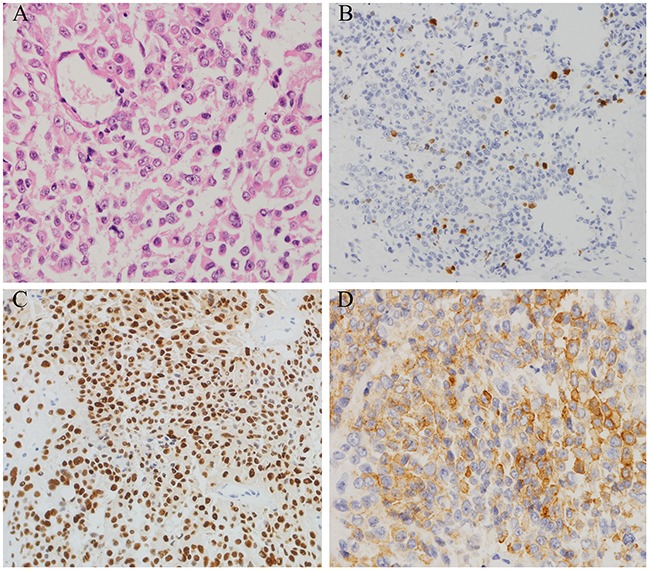
Figure 4. Histopathological findings in resected pituitary tumors from the case two patient.

A. Haematoxylin and eosin (H&E) staining of pituitary tumor (40×); B. The Ki-67 index was greater than 5% (40×); C. Diffuse and strong nuclear p53 staining (arrow) was observed in 90% of tumor cells (40×); D. 90% of tumor cells were positive for ACTH (40×).
