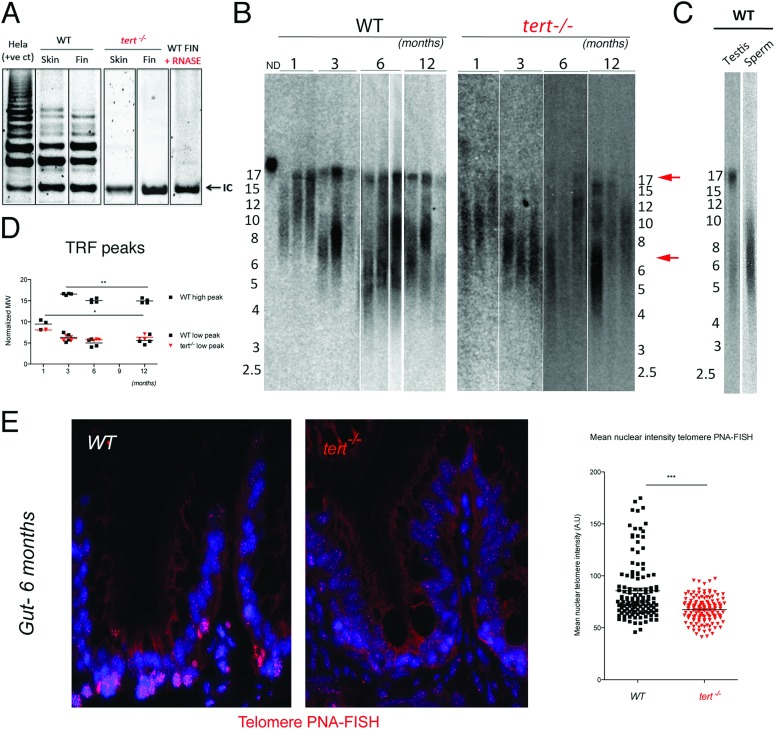
In panel A of Fig 1, a duplication of the tert−/− Skin lane appears where the tert−/− Fin lane should be. Please view the correct Fig 1 here with the correct tert−/− Fin lane shown. Further clarification and copies of the original gels can be found in S1 File.
Fig 1. Telomerase mutant zebrafish have shorter telomeres than WT siblings.
A) Representative image of TRAP assay showing that telomerase is not active in the tert−/− zebrafish, as compared to tert+/+ siblings. Here shown are caudal fin and skin protein extracts. Hela cell extract is shown as positive control. N = 4. B) Representative image of restriction fragment analysis of caudal fin genomic DNA of 3 different individuals at different ages, by southern blot (random primer-labelled telomeric probe (CCCTAA)12 32P-dCTP). tert+/+ Zebrafish have heterogeneous telomeres, with two distinct peaks of different lengths. In tert+/+ the highest peak (∼16 Kb, top red arrow) becomes more distinct after 1 months of age and decreases in length over-time (B and D). The lowest peak of telomere intensity also decreases in length (bottom red arrow, B and D). tert−/− zebrafish have shorter telomeres than tert+/+ siblings in different tissues (see also Figure S1A and S1B), observed by the decrease in length of the higher TRF peak. The shortest TRF peaks accompany those of tert+/+ siblings, and decrease over-time at similar rates. C) Testes fractionation in tert+/+ reveals the two-telomere length populations in whole testes, whereas mature sperm only shows the shorter TRF smear of about 6 Kb, suggesting different telomere lengths in different cells within a tissue. D) TRF mean sizes were calculated as described in [50]. E) Telomere PNA-FISH in 6-month-old gut tissue shows cells with different telomere intensities in the wild type, mainly localizing to the proliferative niche. In contrast tert−/− mutants display cells with less bright and more homogeneous telomere intensity.
Supporting information
(DOCX)
Reference
- 1.Henriques CM, Carneiro MC, Tenente IM, Jacinto A, Ferreira MG (2013) Telomerase Is Required for Zebrafish Lifespan. PLoS Genet 9(1): e1003214 doi:10.1371/journal.pgen.1003214 [DOI] [PMC free article] [PubMed] [Google Scholar]
Associated Data
This section collects any data citations, data availability statements, or supplementary materials included in this article.
Supplementary Materials
(DOCX)


