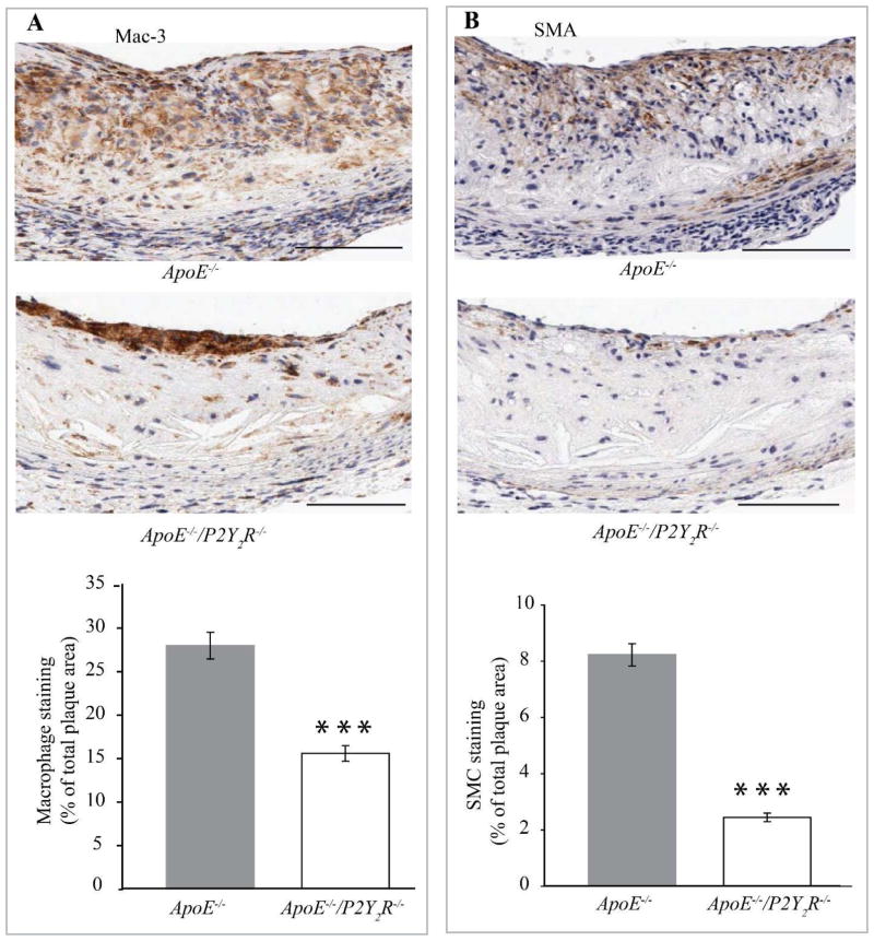
Fig. 2. P2Y2 receptor modulates cellular composition and cellularity of atherosclerotic lesions.
(A and B) Representative images of immunohistological staining of atherosclerotic lesions in the aortic sinus. Adjacent sections were stained with (A) Mac-3 or (B) smooth muscle α-actin antibodies, respectively. The percentage of Mac-3 or smooth muscle α-actin-positive areas in A and B was calculated by dividing the positively stained areas by the total cross-sectional area of the lesion. Bar values are means ± SEM. Five cross sections were evaluated in 12 mice for each genotype. ***p<0.001. Arrows indicate the localization of the staining. Scale bar represents 100 μm.

