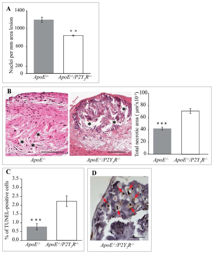
Fig. 3. Reduction of plaque cellularity in ApoE−/−/P2Y2R−/− mice.
(A) Total nuclei were counted in each hematoxylin eosin-stained cross sections (n=5) from 12 mice in each group and normalized to the area lesion. **p<0.01. Scale bar represents 100 μm. (B) Evaluation of plaque necrosis. Representative images of aortic root sections (n=5) from 12 mice of each group genotype were stained with hematoxylin and eosin, and plaque necrosis was quantified. Necrotic areas are indicated with an asterisk. ***p<0.001. Scale bar represents 100 μm. (C) TUNEL-positive nuclei were quantified on lesions from 12 ApoE−/− and 13 ApoE−/−/P2Y2R−/− mice.***p<0.001. (D) High magnification of aortic root cross section from ApoE−/−/P2Y2R−/− mice showing chondrocyte-like cells (asterisk) within large areas of calcification. Scale bar represents 25 μm.

