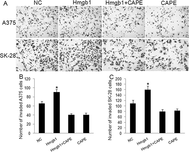
Figure 4. Exogenous Hmgb1 increased invasion in melanoma cells.

A375 and SK-28 cells were treated with Hmgb1 (0.1 μg/ml) with or without 100 μM of NF-κB inhibitor Caffeic Acid Phenethyl Ester (CAPE) for 48 hrs. NC: negative control. Cells were treated with PBS. Hmgb1: cells were treated with Hmgb1 protein (dissolved in PBS). Hmgb1+CAPE: cells were treated with Hmgb1 protein and CAPE. CAPE: cells were only treated with CAPE (dissolved in PBS). A. Representative photographs of cell invasion in A375 and SK-28 cells. B. The number of cell invasion in A375 cells. C. The number of cell invasion in SK-28 cells. *P<0.05 vs. NC group, *P<0.001 vs. other groups. N=4.
