
Figure 3. miR-26a decreased tumor growth in vivo.
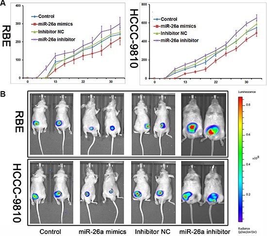
(A) Nude mice were subcutaneously transplanted with cells stably expressed with miR-26a mimics/Inhibitor or control (n = 5). The volume of each tumor was calculated as the length × width2 × 0.5. The unit of y-coordinate was mm3. (B) Mice with established tumors in different groups were imaged every 7 days with the IVIS Lumina II system; the images shown were taken on day 25.