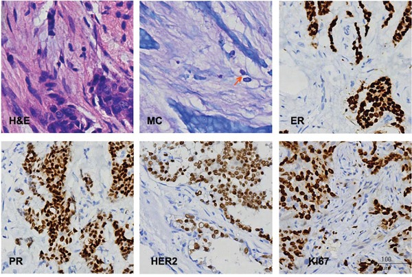
Figure 1. Representative histological staining.

H & E, hematoxylin and eosin staining; MC, mast cells, arrow indicates MC; ER, estrogen receptor; PR, progesterone receptor; HER-2, human epidermal growth factor receptor-2; Ki67, Ki67 nuclear antigen.

H & E, hematoxylin and eosin staining; MC, mast cells, arrow indicates MC; ER, estrogen receptor; PR, progesterone receptor; HER-2, human epidermal growth factor receptor-2; Ki67, Ki67 nuclear antigen.