

Abstract
Protein synthesis underpins much of cell growth and, consequently, cell multiplication. Understanding how proliferating cells commit and progress into the cell cycle requires knowing not only which proteins need to be synthesized, but also what determines their rate of synthesis during cell division.
Keywords: protein synthesis, ribosome biogenesis, ribosome profiling, START, translational control
INTRODUCTION
Experiments with proliferating populations of microbial strains, animal or plant cell lines, have rigorous expectations. Under the same culture conditions, cells ought to have the same properties and composition in every single experiment. The basic “metrics” of proliferating cells remain constant, even after many rounds of cell division 1,2. These metrics include cellular mass and volume, and macromolecular composition 1,3. The constancy of such parameters reflects the fundamental ability of cells to coordinate their growth with their division 1,4,5. Balancing cell growth with cell division determines the overall rates of cell proliferation 4,5,6,7,8. Despite the obvious significance of this phenomenon, how cells manage to coordinate their growth with their division remains largely mysterious.
Proteins are often the most abundant macromolecules in proliferating cells. For example, in steady-state cultures of the budding yeast Saccharomyces cerevisiae, the protein content ranges from 35% to 44% of all macromolecules, depending on culture conditions 3. Furthermore, much of the proteome (>20%) is dedicated to making ribosomes and translation factors, enabling cells to make more proteins 9. On top of that, making ribosomal components and assembling them into functional ribosomes involves a dizzying array of molecular players and cellular processes 10,11,12. Consequently, protein synthesis is viewed as a fundamental measure of cell growth. Decades ago, a founding father of cell cycle studies put it this way: “No sensible interpretation of cell growth can be made without a knowledge of the overall pattern of protein synthesis” 13.
In the following sections, we discuss the interplay of protein synthesis and cell division. Examples of translational control in embryonic and meiotic cell divisions have been covered comprehensively elsewhere 14,15. Here, the focus is on mitotic cell division and specifically on the G1 phase of the cell cycle, when cells commit to a new round of cell division. The examples discussed are mainly, but not exclusively, from the budding yeast S. cerevisiae. The discussion centers on un-perturbed, continuously dividing cells, and the impact of genetic, nutritional or chemical perturbations.
Figure 1. FIGURE 1: Schematic overview of the topics covered in this review.
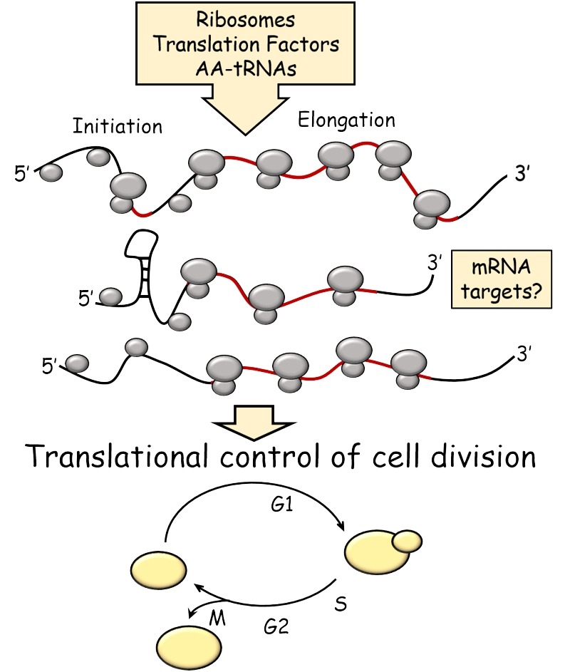
Open reading frames (ORFs) are shown in red.
OVERALL PATTERN OF PROTEIN SYNTHESIS IN THE CELL CYCLE
In animal cells, protein synthesis is much lower in mitosis than in other cell cycle phases 16,17,18. Mechanisms that enable translation of specific mRNAs in animal cells undergoing mitosis have been reviewed elsewhere 15,19. In contrast to the mitotic block in protein synthesis in animal cells, early studies indicated that budding yeast cells synthesize proteins, including ribosomal proteins, continuously during the cell cycle 20,21,22,23. These experiments relied mostly on incorporation of labeled amino acids into polypeptides, which were then visualized after electrophoresis 21,22,23. Hence, those early experiments sampled abundant, constitutively expressed proteins that make up the vast majority of the proteome 9,24,25,26. Obviously, transcriptional waves drive periodic synthesis of hundreds of proteins in the cell cycle 27,28. Nonetheless, the bulk of cellular protein synthesis appears to proceed at an exponentially increasing rate in the cell cycle 21,22. This conclusion was reinforced by monitoring the accumulation of constitutively expressed fluorescent proteins in single cells 29. In addition, continuous monitoring of cell volume supports an exponential mode of increase in the cell cycle 30. Therefore, it appears that budding yeast cells make proteins and grow exponentially. Based on buoyant mass as a metric of cell growth, the same can be said about the growth of diverse types of cells, from bacteria to mouse lymphoblasts, with heavier cells growing faster than lighter cells 31. However, whether or not the growth of animal cells is exponential is still controversial 32,33,34.
An exponential mode of protein synthesis and growth is consistent with the existence of active mechanisms that sense some growth metric, perhaps somehow related to protein synthesis 7. Such mechanisms would enable cells to monitor their growth and commit to a new round of cell division once their growth requirements are met 6,7. As a result, cells that are born small stay longer in the G1 phase, until they grow enough to commit into and initiate a new round of cell division 4,5,6,7,35. In yeast, the point of commitment to a new round of cell division is called START 5. START is marked molecularly by nuclear eviction of the Whi5p repressor 29,36,37, a protein that functions analogously to the retinoblastoma gene product of animal cells 38,39. Once cells pass through START in late G1, they will initiate and complete their division even if they encounter growth limitations 4,5,35. To summarize simply, it seems that the bigger yeast cells get, the faster they make proteins and grow, propelling them to divide. This simple concept raises a series of key questions: What determines the rate of protein synthesis? How can the rate of protein synthesis be altered and what would the effects of such alterations be on the cell cycle? What are the RNA targets of translational control that affect cell cycle progression?
INITIATE TO START?
The rate of synthesis of any given protein depends on not only the concentration but also the translational efficiency of its mRNA. Discrepancies between the two parameters underpin translational control. It is often stated that control of translation in eukaryotic cells is exercised mainly at the initiation step, when ribosomes are recruited to mRNA 19. As discussed in subsequent sections, additional layers of control may also change the rate at which proteins are made. Nonetheless, the initiation step remains a key control point of translation 40,41. Ribosomal recruitment in eukaryotes usually involves recognition of a cap structure at the 5’-end of the mRNA. The small (40S) ribosomal subunit loaded with initiator Met-tRNA and with the contribution of various initiation factors begins scanning the 5’-UTR of the mRNA for an AUG (or near-cognate start codons). In the process, it has to navigate past the secondary structure of the 5’-UTR 42 or initiation codons upstream of the main open reading frame 43. Such features may affect recognition and initiation from the correct start codon 40,41,44.
The earliest genetic evidence for specific cell cycle effects due to translational control was the isolation of budding yeast conditional mutants in what turned out to be translation initiation factors 5. One would expect that cessation of a continuous vital cellular function, such as initiation of translation, would simply arrest each cell at whichever point in the cycle that cell happened to be at the time. In an asynchronously proliferating cell population, this would manifest in a pattern of random arrests along the cell cycle 5. Yet cell division cycle (cdc) genetic screens yielded mutants carrying temperature-sensitive, hypomorphic alleles of translation initiation factors, which did not display a random arrest at their non-permissive temperature. Instead, cells carrying cdc33 (encoding mRNA cap binding protein and translation initiation factor eIF4E 45,46) or cdc63 (encoding the b subunit of translation initiation factor eIF3 47,48) mutations arrest uniformly in the G1 phase of the cell cycle, unable to initiate DNA replication and a new round of cell division 35,46,49,50. A conditional methionyl-tRNA synthetase (mes1) mutant also arrests in the G1 phase of the cell cycle 51. These classical genetic analyses suggested strongly that G1 transit is sensitive to translation initiation, more so than other phases of the cell cycle. This conclusion was strengthened when essential gene function was interrogated with a collection of titratable TetO7 promoter alleles for essential genes 52. In addition to the eIF4E and eIF3b examples mentioned above, Yu et al. showed that inhibiting expression of eIF2a, eIF4A, eIF2b, eIF3i, or eIF1 resulted in G1 arrest in yeast (52; and Table 1). Hence, impairing translation initiation in a number of ways, invariably and specifically also impairs the capacity of cells to initiate a new round of cell division.
Table 1.
Cell cycle phenotypes of loss-of-function mutants in essential genes encoding protein synthesis and ribosome biogenesis factors in S. cerevisiae.
| Systematic name | Standard name/ alias | Function | Cell cycle phenotype | Ref. |
| Translation Initiation Factors | ||||
| YER165W | PAB1 | Poly(A) binding protein | G1 | 136 |
| YJR007W | SUI2 | eIF2α | G1 | 52 |
| YKR059W | TIF1 | eIF4A | G1 | 52 |
| YLR291C | GCD7 | eIF2Bβ | G1 | 52 |
| YMR146C | TIF34 | eIF3i | G1 | 52 |
| YNL244C | SUI1/MOF2 | eIF1 | G1 | 52 |
| YOL139C | CDC33/TIF45 | eIF4E | G1 | 45,46 |
| YOR361C | PRT1/CDC63 | eIF3b | G1 | 47,48 |
| Translation Elongation Factors | ||||
| YLR249W | YEF3 | eEF1Bγ | G2/M, other | 52 |
| tRNA synthetases | ||||
| YGR264C | MES1 | MetRS | G1 | 51 |
| YLL018C | DPS1 | AspRS | G1 | 52 |
| YOR335C | ALA1/CDC64 | AlaRS | G1 | 50 |
| YPL160W | CDC60 | LeuRS | G1 | 50 |
| tRNA | ||||
| tQ(CUG)M | CDC65 | tRNA-Gln | G1 | 137 |
| Ribosome biogenesis and assembly | ||||
| YBL004W | UTP20 | 18S rRNA biogenesis | other | 52 |
| YBR142W | MAK5 | 60S ribosome subunit biogenesis | G1 | 52 |
| YCL054W | SPB1 | AdoMet-dependent methyltransferase | G1 | 52 |
| YCR057C | PWP2/UTP1 | 18S rRNA biogenesis | G1 | 108,138 |
| YDL031W | DBP10 | 40S biogenesis and 35S pre-rRNA processing | G1 | 52 |
| YDL060W | TSR1 | 20S pre-rRNA processing | G1 | 52 |
| YDL148C | NOP14/UTP2 | 18S rRNA biogenesis | G1 | 138 |
| YDL153C | SAS10/UTP3 | 18S rRNA biogenesis | G1, other | 52,138 |
| YDL166C | FAP7 | 20S pre-rRNA processing | G1 | 52 |
| YDR060W | MAK21 | 60S ribosome subunit biogenesis | G1 | 52 |
| YDR091C | RLI1 | Ribosome biogenesis | G1 | 52 |
| YDR324C | UTP4 | 18S rRNA biogenesis | G1 | 138 |
| YDR398W | UTP5 | 18S rRNA biogenesis | G1 | 52,138 |
| YDR449C | UTP6 | 18S rRNA biogenesis | G1 | 138 |
| YER006W | NUG1 | Export of 60S ribosomal subunits from the nucleus | G1 | 52 |
| YER082C | UTP7 | 18S rRNA biogenesis | G1 | 52,138 |
| YER127W | LCP5 | 18S rRNA maturation | G1 | 52 |
| YFL002C | SPB4 | 60S ribosome biogenesis | G1 | 52 |
| YGR090W | UTP22 | 18S rRNA biogenesis | G1 | 52 |
| YGR103W | NOP7 | 60S ribosome subunit biogenesis | G1 | 52 |
| YGR128C | UTP8 | 18S rRNA biogenesis | G1 | 52,138 |
| YGR245C | SDA1 | 60S ribosome biogenesis and actin organization | G1 | 52 |
| YHR072W-A | NOP10 | 18S rRNA maturation | G2/M | 52 |
| YHR085W | IPI1 | 35S pre-rRNA processing | G1 | 52 |
| YHR088W | RPF1 | Export of 60S ribosomal subunits from the nucleus | G1 | 52 |
| YHR089C | GAR1 | Modification and cleavage of the 18S pre-rRNA | other | 52 |
| YHR143W-A | RPC10 | RNA polymerase subunit common to RNA polymerases I, II, and III | G2/M | 52 |
| YHR196W | UTP9 | 18S rRNA biogenesis | G1 | 52,138 |
| YJL033W | HCA4 | 18S rRNA biogenesis | G1 | 52 |
| YJL069C | UTP18 | 18S rRNA biogenesis | G1 | 52 |
| YJL109C | UTP10 | 18S rRNA biogenesis | G1 | 138 |
| YJR002W | MPP10 | 18S rRNA biogenesis | G1, other | 52 |
| YKL009W | MRT4 | Ribosome assembly | G1 | 52 |
| YKL099C | UTP11 | 18S rRNA biogenesis | G1 | 52,138 |
| YKL172W | EBP2 | 25S rRNA maturation | G1 | 52 |
| YLL008W | DRS1 | DEAD-box protein, 60S ribosomal subunits | G1 | 52 |
| YLR002C | NOC3 | 60S ribosome subunit biogenesis | G2/M | 52 |
| YLR009W | RLP24 | 60S ribosome subunit biogenesis | G1 | 52 |
| YLR129W | DIP2 | 18S rRNA biogenesis | G1 | 52,138 |
| YLR167W | RPS31 | Ribosomal protein | G1, other | 52 |
| YLR175W | CBF5 | Pseudouridine synthase | other | 52 |
| YLR186W | EMG1 | Methyltransferase for rRNA | G1 | 52 |
| YLR222C | UTP13 | 18S rRNA biogenesis | G1 | 138 |
| YLR276C | DPB9 | DEAD-box helicase, 27S rRNA processing | G1 | 52 |
| YML093W | UTP14 | 18S rRNA biogenesis | G1 | 52,138 |
| YMR093W | UTP15 | 18S rRNA biogenesis | G1 | 138 |
| YMR128W | ECM16 | DEAD-box helicase, 18S rRNA synthesis | G1 | 52 |
| YMR290C | HAS1 | Helicase, biogenesis of 40S and 60S ribosome subunits | G1 | 52 |
| YNL113W | RPC19 | RNA polymerase subunit common to RNA polymerases I and III | G1 | 52 |
| YNL124W | NAF1 | pre-rRNA processing | G1 | 52 |
| YNL163C | RIA1 | 80S ribosome assembly | G1 | 52 |
| YNL207W | RIO2 | 40S ribosome subunit biogenesis | G1 | 52 |
| YNR038w | DPB6 | DEAD-box helicase | G1 | 52 |
| YNR053C | NOG2 | 60S ribosome subunit biogenesis | G1 | 52 |
| YOL010W | RCL1 | 18S rRNA maturation | G2/M, other | 52 |
| YOR078W | BUD21/UTP16 | 18S rRNA biogenesis | G1 | 138 |
| YOR119C | RIO1 | 40S ribosome subunit biogenesis | other | 52 |
| YOR210W | RPB10 | RNA polymerase subunit common to RNA polymerases I, II, and III | G1 | 52 |
| YOR224C | RPB8 | RNA polymerase subunit common to RNA polymerases I, II, and III | G1 | 52 |
| YOR294W | RRS1 | Export of 60S ribosomal subunits from the nucleus | G1, other | 52 |
| YOR340C | RPA43 | RNA polymerase I subunit | G1 | 52 |
| YOR341W | RPA190 | RNA polymerase I subunit | G1 | 52 |
| YPL012W | RRP12 | Export of ribosomal subunits from the nucleus | G1 | 52 |
| YPL043W | NOP4 | 27S rRNA processing, 60S ribosome subunit biogenesis | G1, other | 52 |
| YPL093W | NOG1 | 60S ribosome subunit biogenesis | G1 | 52 |
| YPL126W | NAN1 | 18S rRNA biogenesis | G1 | 52,138 |
| YPL211W | NIP7 | 60S ribosome subunit biogenesis | G1, other | 52 |
| YPL217C | BMS1 | 40S synthesis and 35S pre-rRNA processing | G1 | 52 |
| YPL266W | DIM1 | 18S rRNA dimethylase | G1 | 52 |
| YPR016C | TIF6/CDC95 | eIF6 | G1 | 52 |
| YPR110C | RPC40 | RNA polymerase subunit common to RNA polymerases I and III | G1 | 52 |
| YPR144C | UTP19 | Maturation and nuclear export of 40S ribosomal subunits | G1 | 52 |
If initiation of translation is important for commitment to division, then signaling pathways that control initiation of division may do so, at least in part, by regulating translation initiation. Mitogenic pathways would be expected to activate translation initiation, while pathways that convey anti-proliferative signals may inhibit translation initiation. The cardinal example for the former case is the Target of Rapamycin (TOR) pathway. How the TOR pathway activates initiation of translation and overall protein synthesis has been reviewed elsewhere 53,54. Loss of TOR function was known to cause G1 arrest in mammals 55,56 and yeast 53,57. Connecting the G1 arrest with the effects of TOR on translation, however, was not obvious. In a landmark paper, it was shown that upon loss of TOR function in yeast, the cause of the G1 arrest was a direct consequence of a block in translation initiation 58. De-repressing translation of the G1 cyclin Cln3p was sufficient to abrogate the G1 arrest of TOR-inhibited yeast cells 58. TOR is not the only mitogenic pathway that activates translation initiation. The RAS/MAPK pathway in animals phosphorylates and increases the activity of eIF4B 40,59. Remarkably, phosphorylation of eIF4B on the same residue is a common output of both the TOR and MAPK pathways 59, underscoring the significance of activating translation initiation for commitment to cell division. Conversely, upon stress or starvation it is not prudent to either initiate cell division or make many proteins. It turns out that phosphorylation of eIF2α is a conserved response from yeast to mammals that inhibits overall translation initiation, and it is an output of anti-mitogenic signals 40,41,53,60.
TRANSLATIONAL TARGETS (?) FOR COMMITMENT TO DIVISION
The above examples suggest that translation initiation goes hand-in-hand with G1 progression and initiation of cell division. By and large, however, they do not answer how this is brought about. What are the relevant proteins important for G1 transit, whose synthesis is sensitive to limitations in translation initiation, and how do these proteins impinge on the machinery of cell division? In the case of the G1 cyclin Cln3p was mentioned above, Hall and colleagues replaced the long 5’-UTR of the yeast CLN3 mRNA with that of UBI4, which is efficiently translated when TOR function is low 58. Cells carrying this non-repressible CLN3 did not arrest in G1 when TOR function was inhibited by rapamycin 58. Similarly, efficient translation of CLN3 enabled G1 arrested cdc33 cells, in which the activity of the eIF4E is impaired (see Table 1), to initiate cell division 61. The Whi3p RNA-binding protein, which sequesters CLN3 mRNA in cytoplasmic foci, may inhibit translation of CLN3 62. There is also a uORF in the 5’-UTR of the CLN3 mRNA 63. We had proposed that the uORF lowers the number of scanning ribosomes that reach the downstream main AUG, especially when the ribosome content of the cell is low in poor media 8,63. As predicted, inactivation of the uORF in CLN3 allowed cells growing in poor medium, with glycerol as the source for carbon, to accelerate completion of START 63. Nitrogen limitation was also reported to repress translation of CLN3 64. In contrast, another study reported that 20 min after amino acid starvation, translation of CLN3 was up-regulated 43. While this later discrepancy may simply reflect the different experimental set-ups, there is overall compelling evidence that synthesis of Cln3p, whose levels control the timing of START 65,66, is regulated at least in part at the translational level. It should be noted, however, that all the examples above rest on comparisons between different conditions: with or without inhibitors of TOR 58,63; mutant vs. wild type 61,62,63; different nutrients 43,63,64. It is important to stress that although CLN3 is a translational target, there is no evidence yet that it is targeted in a periodic manner in the cell cycle, in G1 or any other cell cycle phase.
It has been reported that CLN3 transcription oscillates early in the cell cycle 67,68, but Cln3p protein levels were not evaluated in these studies. Cln3p is very unstable 69, and difficult to detect by immunoblotting. Early studies reported that Cln3p levels do not oscillate in the cell cycle 70. Recently, however, more sensitive approaches from two independent studies showed that Cln3p protein is nearly absent in early G1 cells, but it gradually accumulates as cells approach START (Fig. 9 in 71, and Fig. 10 in 72), without a corresponding increase in the mRNA levels of CLN3. The data from Thorburn et al. 71 and Zapata et al. 72 strongly implicate post-transcriptional mechanisms that control abundance of Cln3p in the cell cycle, perhaps due to control of its synthesis, degradation, or both. Both of these studies relied on centrifugal elutriation to isolate highly synchronous early G1 daughter cells, which were then sampled as they progressed in the cell cycle 71,72. Until analogous studies are performed with alternative synchronization methods, it is formally possible that the results reflect idiosyncrasies of elutriation. Note also that the CLN3 mRNA cannot possibly be the only physiological target of translational control for cell cycle progression. Cells lacking Cln3p are viable 65,66 and they also respond as expected to nutrient limitations, reducing their size 38.
In other systems, the best example of a translational target important for initiation of mitotic cell division is the cyclin-dependent kinase inhibitor p27Kip1 in human cells, whose translation appears to be periodic in the cell cycle, decreasing at the G1/S transition 73,74,75,76,77. Translational control of p27Kip1 is complex, involving both cap-dependent and independent mechanisms 75,77. However, because p27Kip1 synthesis decreases at the G1/S transition, this case of translational control cannot account for the postulated activating role of protein synthesis in triggering cell division. Other reported translational targets include the G1 cyclin D1 in mammals 78,79,80, and the G1/S cyclins E1 81 and E2 82 in mammals, and Cig2 in fission yeast 83 (reviewed in 84). Trans-acting factors influencing translation initiation of these targets include the helicase DDX3 for cyclin E1 81 and Ded1 for Cig2 83. It is not clear, however, if translation of these cyclins is periodic in cycling cells. Alternatively, their translational regulation may be an output of a continuous process that affects their overall levels. The levels of these cyclins may oscillate in the cell cycle for other reasons, such as mechanisms that control mRNA levels and protein degradation. Overall, there is a critical gap in our understanding of the role of translational control in mitotic cell cycle progression, especially in G1 progression and commitment to division. There have not been any studies that directly and systematically looked for mRNAs that are translated differentially in the G1 phase, in cycling, un-perturbed cells.
START ON CYCLOHEXIMIDE
After the initiation step, the rate of translation depends on the concentration and activity of translating ribosomes that elongate the nascent polypeptides. If elongation of protein synthesis is inhibited, then what are the consequences on cell cycle progression? This question was first tackled pharmacologically, monitoring cell cycle progression in the presence of varying doses of cycloheximide 35,85,87,87. Cycloheximide inhibits the translocation step in eukaryotic 80S ribosomes, blocking translational elongation 88. Increasing doses of cycloheximide increase the population doubling time, mostly because cells spend more time in the G1 phase of the cell cycle 35,85. Cycloheximide also affects size homeostasis. In budding yeast, cycloheximide reduces the newborn cell size 35,85 and the rate at which cells increase in size 89. It also increases the critical size threshold for START 85,89. These changes account for the increase in the duration of the G1 phase upon treatment with cycloheximide. The effects of cycloheximide support the notion that a critical rate of protein synthesis is required for G1 transit and completion of START in budding yeast 86 and animal cells 90,91. The reports that interrogated cycloheximide’s effects on G1 progression have been influential. They have often been taken to imply a requirement for the continuous synthesis of unstable protein(s), whose rate of synthesis parallels overall protein synthesis rates and cell growth. Identifying those proteins and how they affect the cell division machinery would then hold the promise of explaining molecularly how cells couple their growth with their division 90,91. Various such candidate proteins have been proposed over the years. However, there is no report of a protein whose levels increase due to cell cycle-dependent translational control, as cycling cells approach START 7.
RIBOSOME MAKES PROTEIN MAKES CELLS?
Ribosomes are the complex macromolecular machines that catalyze protein synthesis. Hence, changing the concentration of functional ribosomes in the cell is expected to change the overall rates of protein synthesis.
Bacterial cells grown under conditions that favor fast growth and proliferation (i.e., "rich" media) have more ribosomes than those propagated in "poor" media 92. These observations suggest that the rate of protein synthesis in bacteria is controlled mostly by ribosome numbers 93. Although growth rate does not seem to affect significantly the fraction of active ribosomes in the cell (≈80%) or their activity 94, recent observations suggest that even in rich media bacterial ribosomes do not function at maximal elongation rates 95. Hence, translation rates may be adjusted to proliferation rates by means other than ribosome content.
In budding yeast, although protein synthesis rates decrease with decreasing growth rates, there is no proportional decrease in the number of ribosomes per cell 96,97. For example, a 10-fold drop in growth rate is only accompanied by a 2-fold drop in the number of ribosomes per cell (see Table 4 in 96). On the other hand, synthesis of ribosomal components is inhibited as cells begin to exhaust the available nutrients and prepare to enter stationary phase 98, or upon amino acid starvation 99. Apparently, both the activity and the number of ribosomes may be affected in yeast as a function of growth rate.
In cycling, unperturbed yeast cells, synthesis of ribosomal components is not cell cycle dependent 10,22,23. Interfering with ribosome biogenesis, however, affects cell cycle progression dramatically. In yeast, 59 of a total of 78 ribosomal proteins of cytoplasmic ribosomes are encoded by pairs of very similar or identical paralogous genes 100. Mutants carrying single deletions of those ribosomal protein genes are usually viable 101. Many of these deletion strains have a small overall cell size 102,103. A small overall cell size is also characteristic of cells lacking Sfp1p, a transcriptional activator of ribosome biosynthesis and ribosomal protein genes 89. Cells lacking Sfp1p are born very small 104, and their growth rate is about half of that of wild type cells 71,104. As a result, the duration of the G1 phase of the cell cycle is greatly increased in sfp1Δ cells 71,89,104. Still, the smaller critical size threshold of sfp1Δ cells and the smaller overall size of ribosomal protein mutants led Tyers and colleagues to propose that ribosome biogenesis inhibits START in wild type cells 6,89. In this model, ribosome biogenesis sets the critical size threshold for START, while translation rates of functional ribosomes enable the cells to pass that threshold 6,89. This model appears paradoxical and counterintuitive, especially since in animals ribosome biosynthesis is thought to promote cell proliferation and cancer 105. But another finding, that ribosomal proteins may function as haploinsufficient tumor suppressors in animals, appeared to offer support for a negative role of ribosome biogenesis in cell division 106. However, it was subsequently reported that such effects may be due to cell non-autonomous routes 107. Furthermore, the following observations argue against the notion that ribosome biogenesis has a general inhibitory role for the initiation of division: First, inhibition of ribosome maturation delays START 108. Second, despite their small cell size, most ribosomal protein mutants have a longer G1 phase 104,109,110. Third, loss-of-function of the vast majority of essential ribosome biogenesis factors leads to G1 arrest (see Table 1 and 52). The most straightforward interpretation of the evidence outlined above is that ribosome biogenesis promotes initiation of cell division in budding yeast.
Even when there is a G1 delay due to loss of a ribosomal protein, G1 variables such as birth and critical size of cells lacking individual ribosomal proteins are not uniform, differing qualitatively and quantitatively 104,109,111. Furthermore, although a delay in G1 is the most common phenotype upon loss of a ribosomal protein, this phenotype is not universal. In several cases, there is no cell cycle phenotype, or a G2/M block is observed instead 104,110. The basis of all those differences in ribosomal protein mutant phenotypes related to the cell cycle is not clear. Do they reflect specialized translational roles of some ribosomes? Are overall translation rates affected? Is the concentration of ribosomes, their composition, their activity, localization in the cell (or any combination of the above) that is affected? Examples of specialized ribosomal functions abound 112,113,114,115, leading some to speculate on the existence of a “ribosome code” 116. There are even cases of extraribosomal roles for ribosomal proteins 117. Regardless of the answers to the above questions, it is imperative that the relevant mRNA substrates affected in each ribosomal protein mutant must be identified, to understand how cell cycle progression might be impacted. However, as is the case for wild type cells (see INITIATE TO START? above), there are no systematic surveys of mRNAs that are translated differentially in the cell cycle in ribosomal protein mutants.
THE OTHER RIBOSOME SUBSTRATES: tRNAS
For the ribosome to elongate at its maximal rate, all its substrates must be present at saturating concentrations. This includes the aminoacyl-tRNAs and the various elongation factors 95. Inhibition of elongation factor eEF2 may be one way that protein synthesis is inhibited in animal cells in mitosis 118. However, unlike the numerous examples of perturbations in translation initiation factors that lead to G1 arrest in yeast (Table 1), there is very little analogous evidence for translation elongation factors affecting cell cycle progression. The only reported example is from a large survey of essential genes, reporting that blocking expression of eEF1Bγ in yeast leads to cell cycle arrest in mitosis (52, see Table 1).
Translation elongation rates may be affected by several parameters 119, including the supply and demand for each tRNA. The genetic code is essentially universal. One and the same codon does not code for different amino acids in different organisms. The code, however, is also degenerate. Most amino acids can be specified by more than one codon. A decades-old speculation has been that codons for an amino acid that are used more frequently in an organism would be translated more rapidly than codons for the same amino acid that are rarely used 120. Differences in codon usage were difficult to test until the advent of ribosome profiling. Sequencing ribosome-protected mRNA fragments allows estimates of ribosome residency on individual codons 43. Several analyses that included the original ribosome profiling datasets in yeast 43 have examined various parameters, including codon usage, and reached contradictory conclusions 42,119,121,122,123,124,125,126. Nonetheless, common codons may be translated faster than rare ones in yeast 127. Modulating which codon is used and the effective concentration of its corresponding tRNA may be a way to influence translation elongation rates both within an mRNA, and across the transcriptome 128. Surveying codon usage across each codon and tRNA availability revealed that in highly expressed mRNAs rare codons are more prevalent in the first 30-50 codons, slowing down and crowding ribosomes along that stretch 129. It was proposed that this slow “ramp” is a mechanism that promotes faster translation downstream, by alleviating ribosome “traffic jams” along the mRNA 129. Furthermore, it turns out that once a particular codon has been used, it will also be used more frequently whenever the same amino acid is encoded downstream in that mRNA 130. Based on these observations, it was theorized that tRNAs do not diffuse away from the ribosome once they are expelled from it. Instead, the same tRNA for a codon specifying the repeating amino acid is re-charged and channeled for re-use, enabling translation to proceed faster than tRNA diffusion 130.
The examples outlined above illustrate how alterations in the supply and demand for each tRNA could impact translation rates. The pertinent question for this review, however, is whether such mechanisms could lead to translational control during the cell cycle. Remarkably, a recent study proposed exactly that kind of regulation, based on codon usage 131. Optimal codon usage is more prevalent in mRNAs expressed in the G1 phase of the cell cycle, implying that the corresponding mRNAs are translated more efficiently 131. Interestingly, it has been known for decades that loss-of-function mutations in several tRNA synthetases, and even in a tRNA gene, lead to G1 arrest in yeast (see Table 1). Optimizing translation elongation rates in G1 through codon usage is an exciting possibility. But a correlation is not causation, and the above predictions must await experimental validation, measuring the translational efficiencies of the putative mRNA targets in cycling cells. On a cautionary note, a recent study in mouse embryonic stem cells reported that translation elongation rates were not only independent of codon usage, but also very similar across different mRNAs 132. Likewise, another study in yeast reported that the rate of translation elongation and translational efficiency were not affected by tRNA abundance, and codon translation rates were not correlated with codon bias 133. Hence, these issues remain controversial and there might be some time before the dust settles.
DIVIDE TO TRANSLATE?
Implicit in the discussion of all the examples mentioned above is the notion that cell growth and protein synthesis drive cell cycle progression, not the other way around. This is generally the case, established in classic and particularly lucid experiments by Hartwell and colleagues 4,5,35. However, there are also some changes in the pattern of growth once cell cycle progression is blocked 134,135. Does that mean that progressing in the cell cycle might also affect the translational control of specific mRNAs? The answer is a resounding yes. Ruggero and colleagues examined by ribosome profiling human cells arrested by thymidine block (G1 and S phases) and nocodazole treatment (G2 phase) in the cell cycle, reporting extensive translational control of numerous mRNAs 17. Interestingly, this work revealed functional clusters of co-regulated mRNAs. Translational control may be used to coordinate expression of specific cellular machines and processes 17. These experiments are very important, answering how cell division controls translation. However, they do not answer how translation controls cell division.
OUTLOOK
The role of protein synthesis and translational control has a long history in the cell cycle field. The pioneering experiments of decades ago were incisive, but also largely descriptive. Since then, progress has been incremental and focused on a limited number of putative mRNA translational targets. Identifying all the mRNAs that are under periodic translational control in cycling cells is an obvious and necessary goal. Given the transformative methodologies now available and the current pace of progress, it is only a matter of time before we know how protein synthesis drives cell cycle progression.
Funding Statement
Work in the authors’ laboratories has been supported with grants from the National Science Foundation (MCB-0818248 to M.P.) and the National Institutes of Health (GM58770 to R.A.). The open access publishing fees for this article have been covered by the Texas A&M University Online Access to Knowledge (OAK) Fund, supported by the University Libraries and the Office of the Vice President for Research.
References
- 1.Neidhardt FC, Ingraham JL, Schaechter M. Sunderland, Massachusetts.: Sinauer Associates, Inc; 1990. Physiology of the bacterial cell. [Google Scholar]
- 2.Meyer HP, Kappeli O, Fiechter A. Growth control in microbial cultures. Annual review of microbiology. 1985;39:299–319. doi: 10.1146/annurev.mi.39.100185.001503. [DOI] [PubMed] [Google Scholar]
- 3.Lange HC, Heijnen JJ. Statistical reconciliation of the elemental and molecular biomass composition of Saccharomyces cerevisiae. Biotechnology and bioengineering. 2001;75(3):334–344. doi: 10.1002/bit.10054. [DOI] [PubMed] [Google Scholar]
- 4.Johnston GC, Pringle JR, Hartwell LH. Coordination of growth with cell division in the yeast Saccharomyces cerevisiae. Experimental cell research. 1977;105(1):79–98. doi: 10.1016/0014-4827(77)90154-9. [DOI] [PubMed] [Google Scholar]
- 5.Pringle JR, Hartwell LH. The Molecular and Cellular Biology of the Yeast Saccharomyces. Cold Spring Harbor Laboratory Press; 1981. The Saccharomyces cerevisiae Cell Cycle. pp. 97–142. [Google Scholar]
- 6.Jorgensen P, Tyers M. How cells coordinate growth and division. Current biology. 2004;14(23):R1014–R1027. doi: 10.1016/j.cub.2004.11.027. [DOI] [PubMed] [Google Scholar]
- 7.Turner JJ, Ewald JC, Skotheim JM. Cell size control in yeast. Current biology. 2012;22(9):R350–R359. doi: 10.1016/j.cub.2012.02.041. [DOI] [PMC free article] [PubMed] [Google Scholar]
- 8.Polymenis M, Schmidt EV. Coordination of cell growth with cell division. Current opinion in genetics & development. 1999;9(1):76–80. doi: 10.1016/S0959-437X(99)80011-2. [DOI] [PubMed] [Google Scholar]
- 9.Liebermeister W, Noor E, Flamholz A, Davidi D, Bernhardt J, Milo R. Visual account of protein investment in cellular functions. Proceedings of the National Academy of Sciences of the United States of America. 2014;111(23):8488–8493. doi: 10.1073/pnas.1314810111. [DOI] [PMC free article] [PubMed] [Google Scholar]
- 10.Warner JR. The economics of ribosome biosynthesis in yeast. Trends in biochemical sciences. 1999;24(11):437–440. doi: 10.1016/S0968-0004(99)01460-7. [DOI] [PubMed] [Google Scholar]
- 11.Fatica A, Tollervey D. Making ribosomes. Current opinion in cell biology. 2002;14(3):313–318. doi: 10.1016/S0955-0674(02)00336-8. [DOI] [PubMed] [Google Scholar]
- 12.Venema J, Tollervey D. Ribosome synthesis in Saccharomyces cerevisiae. Annual review of genetics. 1999;33(1):261–311. doi: 10.1146/annurev.genet.33.1.261. [DOI] [PubMed] [Google Scholar]
- 13.Mitchison JM. The Biology of the Cell Cycle. Cambridge University Press; 1971. Cell Growth and Protein Synthesis. p. 129. [Google Scholar]
- 14.Brar GA, Yassour M, Friedman N, Regev A, Ingolia NT, Weissman JS. High-resolution view of the yeast meiotic program revealed by ribosome profiling. Science. 2012;335(6068):552–557. doi: 10.1126/science.1215110. [DOI] [PMC free article] [PubMed] [Google Scholar]
- 15.Kronja I, Orr-Weaver TL. Translational regulation of the cell cycle: when, where, how and why? Philosophical transactions of the Royal Society of London Series B, Biological sciences. 2011;366(1584):3638–3652. doi: 10.1098/rstb.2011.0084. [DOI] [PMC free article] [PubMed] [Google Scholar]
- 16.Prescott D, Bender M. Synthesis of RNA and protein during mitosis in mammalian tissue culture cells. Experimental cell research. 1962;26(2):260–268. doi: 10.1016/0014-4827(62)90176-3. [DOI] [PubMed] [Google Scholar]
- 17.Stumpf CR, Moreno MV, Olshen AB, Taylor BS, Ruggero D. The translational landscape of the mammalian cell cycle. Molecular cell. 2013;52(4):574–582. doi: 10.1016/j.molcel.2013.09.018. [DOI] [PMC free article] [PubMed] [Google Scholar]
- 18.Scharff MD, Robbins E. Polyribosome disaggregation during metaphase. Science. 1966;151(3713):992. doi: 10.1126/science.151.3713.992. [DOI] [PubMed] [Google Scholar]
- 19.Pyronnet S, Sonenberg N. Cell-cycle-dependent translational control. Current opinion in genetics & development. 2001;11(1):13–18. doi: 10.1016/S0959-437X(00)00150-7. [DOI] [PubMed] [Google Scholar]
- 20.Williamson D, Scopes A. Protein synthesis and nitrogen uptake in synchronously dividing cultures of Saccharomyces cerevisiae. Journal of the Institute of Brewing. 1961;67(1):39–42. doi: 10.1002/j.2050-0416.1961.tb01755.x. [DOI] [Google Scholar]
- 21.Elliott SG, McLaughlin CS. Rate of macromolecular synthesis through the cell cycle of the yeast Saccharomyces cerevisiae. Proceedings of the National Academy of Sciences of the United States of America. 1978;75(9):4384–4388. doi: 10.1073/pnas.75.9.4384. [DOI] [PMC free article] [PubMed] [Google Scholar]
- 22.Elliott SG, Warner JR, McLaughlin CS. Synthesis of ribosomal proteins during the cell cycle of the yeast Saccharomyces cerevisiae. Journal of bacteriology. 1979;137(2):1048–1050. doi: 10.1128/MCB.2.2.117. [DOI] [PMC free article] [PubMed] [Google Scholar]
- 23.Shulman RW, Hartwell LH, Warner JR. Synthesis of ribosomal proteins during the yeast cell cycle. Journal of molecular biology. 1973;73(4):513–525. doi: 10.1016/0022-2836(73)90097-1. [DOI] [PubMed] [Google Scholar]
- 24.Nagaraj N, Kulak NA, Cox J, Neuhauser N, Mayr K, Hoerning O, Vorm O, Mann M. System-wide perturbation analysis with nearly complete coverage of the yeast proteome by single-shot ultra HPLC runs on a bench top Orbitrap. Molecular & cellular proteomics. 2012;11(3):M111.013722. doi: 10.1074/mcp.M111.013722. [DOI] [PMC free article] [PubMed] [Google Scholar]
- 25.Lu P, Vogel C, Wang R, Yao X, Marcotte EM. Absolute protein expression profiling estimates the relative contributions of transcriptional and translational regulation. Nature biotechnology. 2007;25(1):117–124. doi: 10.1038/nbt1270. [DOI] [PubMed] [Google Scholar]
- 26.Futcher B, Latter G, Monardo P, McLaughlin C, Garrels J. A sampling of the yeast proteome. Molecular and cellular biology. 1999;19(11):7357–7368. doi: 10.1128/mcb.19.11.7357. [DOI] [PMC free article] [PubMed] [Google Scholar]
- 27.Spellman PT, Sherlock G, Zhang MQ, Iyer VR, Anders K, Eisen MB, Brown PO, Botstein D, Futcher B. Comprehensive identification of cell cycle-regulated genes of the yeast Saccharomyces cerevisiae by microarray hybridization. Molecular biology of the cell. 1998;9(12):3273–3297. doi: 10.1091/mbc.9.12.3273. [DOI] [PMC free article] [PubMed] [Google Scholar]
- 28.Futcher B. Transcriptional regulatory networks and the yeast cell cycle. Current opinion in cell biology. 2002;14(6):676–683. doi: 10.1016/s0955-0674(02)00391-5. [DOI] [PubMed] [Google Scholar]
- 29.Di Talia S, Skotheim JM, Bean JM, Siggia ED, Cross FR. The effects of molecular noise and size control on variability in the budding yeast cell cycle. Nature. 2007;448(7156):947–951. doi: 10.1038/nature06072. [DOI] [PubMed] [Google Scholar]
- 30.Bryan AK, Engler A, Gulati A, Manalis SR. Continuous and long-term volume measurements with a commercial Coulter counter. PloS one. 2012;7(1):e29866. doi: 10.1371/journal.pone.0029866. [DOI] [PMC free article] [PubMed] [Google Scholar]
- 31.Godin M, Delgado FF, Son S, Grover WH, Bryan AK, Tzur A, Jorgensen P, Payer K, Grossman AD, Kirschner MW. Using buoyant mass to measure the growth of single cells. Nature methods. 2010;7(5):387–390. doi: 10.1038/nmeth.1452. [DOI] [PMC free article] [PubMed] [Google Scholar]
- 32.Conlon I, Raff M. Differences in the way a mammalian cell and yeast cells coordinate cell growth and cell-cycle progression. Journal of biology. 2003;2(1):7. doi: 10.1186/1475-4924-2-7. [DOI] [PMC free article] [PubMed] [Google Scholar]
- 33.Echave P, Conlon IJ, Lloyd AC. Cell size regulation in mammalian cells. Cell cycle. 2007;6(2):218–224. doi: 10.4161/cc.6.2.3744. [DOI] [PubMed] [Google Scholar]
- 34.Grewal SS, Edgar BA. Controlling cell division in yeast and animals: does size matter? Journal of biology. 2003;2(1):5. doi: 10.1186/1475-4924-2-5. [DOI] [PMC free article] [PubMed] [Google Scholar]
- 35.Hartwell LH, Unger MW. Unequal division in Saccharomyces cerevisiae and its implications for the control of cell division. The Journal of cell biology. 1977;75(2 Pt 1):422–435. doi: 10.1083/jcb.75.2.422. [DOI] [PMC free article] [PubMed] [Google Scholar]
- 36.Eser U, Falleur-Fettig M, Johnson A, Skotheim JM. Commitment to a cellular transition precedes genome-wide transcriptional change. Molecular cell. 2011;43(4):515–527. doi: 10.1016/j.molcel.2011.06.024. [DOI] [PMC free article] [PubMed] [Google Scholar]
- 37.Doncic A, Falleur-Fettig M, Skotheim Jan M. Distinct Interactions Select and Maintain a Specific Cell Fate. Molecular cell. 2011;43(4):528–539. doi: 10.1016/j.molcel.2011.06.025. [DOI] [PMC free article] [PubMed] [Google Scholar]
- 38.Costanzo M, Nishikawa JL, Tang X, Millman JS, Schub O, Breitkreuz K, Dewar D, Rupes I, Andrews B, Tyers M. CDK activity antagonizes Whi5, an inhibitor of G1/S transcription in yeast. Cell. 2004;117(7):899–913. doi: 10.1016/j.cell.2004.05.024. [DOI] [PubMed] [Google Scholar]
- 39.de Bruin RA, McDonald WH, Kalashnikova TI, Yates 3rd J, Wittenberg C. Cln3 activates G1-specific transcription via phosphorylation of the SBF bound repressor Whi5. Cell. 2004;117(7):887–898. doi: 10.1016/j.cell.2004.05.025. [DOI] [PubMed] [Google Scholar]
- 40.Sonenberg N, Hinnebusch AG. Regulation of translation initiation in eukaryotes: mechanisms and biological targets. Cell. 2009;136(4):731–745. doi: 10.1016/j.cell.2009.01.042. [DOI] [PMC free article] [PubMed] [Google Scholar]
- 41.Hershey JW, Sonenberg N, Mathews MB. Principles of translational control: an overview. Cold Spring Harbor perspectives in biology. 2012;4(12) doi: 10.1101/cshperspect.a011528. [DOI] [PMC free article] [PubMed] [Google Scholar]
- 42.Kertesz M, Wan Y, Mazor E, Rinn JL, Nutter RC, Chang HY, Segal E. Genome-wide measurement of RNA secondary structure in yeast. Nature. 2010;467(7311):103–107. doi: 10.1038/nature09322. [DOI] [PMC free article] [PubMed] [Google Scholar]
- 43.Ingolia NT, Ghaemmaghami S, Newman JR, Weissman JS. Genome-wide analysis in vivo of translation with nucleotide resolution using ribosome profiling. Science. 2009;324(5924):218–223. doi: 10.1126/science.1168978. [DOI] [PMC free article] [PubMed] [Google Scholar]
- 44.Hood HM, Neafsey DE, Galagan J, Sachs MS. Evolutionary roles of upstream open reading frames in mediating gene regulation in fungi. Annual review of microbiology. 2009;63:385–409. doi: 10.1146/annurev.micro.62.081307.162835. [DOI] [PubMed] [Google Scholar]
- 45.Altmann M, Trachsel H. Altered mRNA cap recognition activity of initiation factor 4E in the yeast cell cycle division mutant cdc33. Nucleic acids research. 1989;17(15):5923–5931. doi: 10.1093/nar/17.15.5923. [DOI] [PMC free article] [PubMed] [Google Scholar]
- 46.Brenner C, Nakayama N, Goebl M, Tanaka K, Toh-e A, Matsumoto K. CDC33 encodes mRNA cap-binding protein eIF-4E of Saccharomyces cerevisiae. Molecular and cellular biology. 1988;8(8):3556–3559. doi: 10.1128/MCB.8.8.3556. [DOI] [PMC free article] [PubMed] [Google Scholar]
- 47.Naranda T, MacMillan SE, Hershey JW. Purified yeast translational initiation factor eIF-3 is an RNA-binding protein complex that contains the PRT1 protein. The Journal of biological chemistry. 1994;269(51):32286–32292. [PubMed] [Google Scholar]
- 48.Keierleber C, Wittekind M, Qin SL, McLaughlin CS. Isolation and characterization of PRT1, a gene required for the initiation of protein biosynthesis in Saccharomyces cerevisiae. Molecular and cellular biology. 1986;6(12):4419–4424. doi: 10.1128/MCB.6.12.4419. [DOI] [PMC free article] [PubMed] [Google Scholar]
- 49.Hanic-Joyce PJ, Johnston GC, Singer RA. Regulated arrest of cell proliferation mediated by yeast prt1 mutations. Experimental cell research. 1987;172(1):134–145. doi: 10.1016/0014-4827(87)90100-5. [DOI] [PubMed] [Google Scholar]
- 50.Bedard DP, Johnston GC, Singer RA. New mutations in the yeast Saccharomyces cerevisiae affecting completion of "start". Current genetics. 1981;4(3):205–214. doi: 10.1007/BF00420500. [DOI] [PubMed] [Google Scholar]
- 51.Unger MW, Hartwell LH. Control of cell division in Saccharomyces cerevisiae by methionyl-tRNA. Proceedings of the National Academy of Sciences of the United States of America. 1976;73(5):1664–1668. doi: 10.1073/pnas.73.5.1664. [DOI] [PMC free article] [PubMed] [Google Scholar]
- 52.Yu L, Pena Castillo L, Mnaimneh S, Hughes TR, Brown GW. A survey of essential gene function in the yeast cell division cycle. Molecular biology of the cell. 2006;17(11):4736–4747. doi: 10.1091/mbc.E06-04-0368. [DOI] [PMC free article] [PubMed] [Google Scholar]
- 53.Loewith R, Hall MN. Target of rapamycin (TOR) in nutrient signaling and growth control. Genetics. 2011;189(4):1177–1201. doi: 10.1534/genetics.111.133363. [DOI] [PMC free article] [PubMed] [Google Scholar]
- 54.Ma XM, Blenis J. Molecular mechanisms of mTOR-mediated translational control. Nature reviews Molecular cell biology. 2009;10(5):307–318. doi: 10.1038/nrm2672. [DOI] [PubMed] [Google Scholar]
- 55.Wicker LS, Boltz RC, Matt V, Nichols EA, Peterson LB, Sigal NH. Suppression of B cell activation by cyclosporin A, FK506 and rapamycin. European journal of immunology. 1990;20(10):2277–2283. doi: 10.1002/eji.1830201017. [DOI] [PubMed] [Google Scholar]
- 56.Zoncu R, Efeyan A, Sabatini DM. mTOR: from growth signal integration to cancer, diabetes and ageing. Nature reviews Molecular cell biology. 2010;12(1):21–35. doi: 10.1038/nrm3025. [DOI] [PMC free article] [PubMed] [Google Scholar]
- 57.Heitman J, Movva NR, Hall MN. Targets for cell cycle arrest by the immunosuppressant rapamycin in yeast. Science. 1991;253(5022):905–909. doi: 10.1126/science.1715094. [DOI] [PubMed] [Google Scholar]
- 58.Barbet NC, Schneider U, Helliwell SB, Stansfield I, Tuite MF, Hall MN. TOR controls translation initiation and early G1 progression in yeast. Molecular biology of the cell. 1996;7(1):25–42. doi: 10.1091/mbc.7.1.25. [DOI] [PMC free article] [PubMed] [Google Scholar]
- 59.Shahbazian D, Roux PP, Mieulet V, Cohen MS, Raught B, Taunton J, Hershey JW, Blenis J, Pende M, Sonenberg N. The mTOR/PI3K and MAPK pathways converge on eIF4B to control its phosphorylation and activity. The EMBO journal. 2006;25(12):2781–2791. doi: 10.1038/sj.emboj.7601166. [DOI] [PMC free article] [PubMed] [Google Scholar]
- 60.Cherkasova VA, Hinnebusch AG. Translational control by TOR and TAP42 through dephosphorylation of eIF2alpha kinase GCN2. Genes & development. 2003;17(7):859–872. doi: 10.1101/gad.1069003. [DOI] [PMC free article] [PubMed] [Google Scholar]
- 61.Danaie P, Altmann M, Hall M, Trachsel H, Helliwell S. CLN3 expression is sufficient to restore G1-to-S-phase progression in Saccharomyces cerevisiae mutants defective in translation initiation factor eIF4E. Biochem J. 1999;340:135–141. doi: 10.1042/0264-6021:3400135. [DOI] [PMC free article] [PubMed] [Google Scholar]
- 62.Cai Y, Futcher B. Effects of the yeast RNA-binding protein Whi3 on the half-life and abundance of CLN3 mRNA and other targets. PloS one. 2013;8(12):e84630. doi: 10.1371/journal.pone.0084630. [DOI] [PMC free article] [PubMed] [Google Scholar]
- 63.Polymenis M, Schmidt EV. Coupling of cell division to cell growth by translational control of the G1 cyclin CLN3 in yeast. Genes & development. 1997;11(19):2522–2531. doi: 10.1101/gad.11.19.2522. [DOI] [PMC free article] [PubMed] [Google Scholar]
- 64.Gallego C, Garí E, Colomina N, Herrero E, Aldea M. The Cln3 cyclin is down‐regulated by translational repression and degradation during the G1 arrest caused by nitrogen deprivation in budding yeast. The EMBO journal. 1997;16(23):7196–7206. doi: 10.1093/emboj/16.23.7196. [DOI] [PMC free article] [PubMed] [Google Scholar]
- 65.Nash R, Tokiwa G, Anand S, Erickson K, Futcher AB. The WHI1+ gene of Saccharomyces cerevisiae tethers cell division to cell size and is a cyclin homolog. The EMBO journal. 1988;7(13):4335–4346. doi: 10.1002/j.1460-2075.1988.tb03332.x. [DOI] [PMC free article] [PubMed] [Google Scholar]
- 66.Cross FR. DAF1, a mutant gene affecting size control, pheromone arrest, and cell cycle kinetics of Saccharomyces cerevisiae. Molecular and cellular biology. 1988;8(11):4675–4684. doi: 10.1128/MCB.8.11.4675. [DOI] [PMC free article] [PubMed] [Google Scholar]
- 67.Di Talia S, Wang H, Skotheim JM, Rosebrock AP, Futcher B, Cross FR. Daughter-Specific Transcription Factors Regulate Cell Size Control in Budding Yeast. PLoS biology. 2009;7(10):e1000221. doi: 10.1371/journal.pbio.1000221. [DOI] [PMC free article] [PubMed] [Google Scholar]
- 68.MacKay VL, Mai B, Waters L, Breeden LL. Early Cell Cycle Box-Mediated Transcription ofCLN3 and SWI4 Contributes to the Proper Timing of the G1-to-S Transition in Budding Yeast. Molecular and cellular biology. 2001;21(13):4140–4148. doi: 10.1128/mcb.21.13.4140-4148.2001. [DOI] [PMC free article] [PubMed] [Google Scholar]
- 69.Tyers M, Tokiwa G, Nash R, Futcher B. The Cln3-Cdc28 kinase complex of S. cerevisiae is regulated by proteolysis and phosphorylation. The EMBO journal. 1992;11(5):1773–1784. doi: 10.1002/j.1460-2075.1992.tb05229.x. [DOI] [PMC free article] [PubMed] [Google Scholar]
- 70.Tyers M, Tokiwa G, Futcher B. Comparison of the Saccharomyces cerevisiae G1 cyclins: Cln3 may be an upstream activator of Cln1, Cln2 and other cyclins. The EMBO journal. 1993;12(5):1955–1968. doi: 10.1002/j.1460-2075.1993.tb05845.x. [DOI] [PMC free article] [PubMed] [Google Scholar]
- 71.Thorburn RR, Gonzalez C, Brar GA, Christen S, Carlile TM, Ingolia NT, Sauer U, Weissman JS, Amon A. Aneuploid yeast strains exhibit defects in cell growth and passage through START. Molecular biology of the cell. 2013;24(9):1274–1289. doi: 10.1091/mbc.E12-07-0520. [DOI] [PMC free article] [PubMed] [Google Scholar]
- 72.Zapata J, Dephoure N, MacDonough T, Yu Y, Parnell EJ, Mooring M, Gygi SP, Stillman DJ, Kellogg DR. PP2ARts1 is a master regulator of pathways that control cell size. The Journal of cell biology. 2014;204(3):359–376. doi: 10.1083/jcb.201309119. [DOI] [PMC free article] [PubMed] [Google Scholar]
- 73.Gopfert U, Kullmann M, Hengst L. Cell cycle-dependent translation of p27 involves a responsive element in its 5'-UTR that overlaps with a uORF. Human molecular genetics. 2003;12(14):1767–1779. doi: 10.1093/hmg/ddg177. [DOI] [PubMed] [Google Scholar]
- 74.Hengst L, Reed SI. Translational control of p27Kip1 accumulation during the cell cycle. Science. 1996;271(5257):1861–1864. doi: 10.1126/science.271.5257.1861. [DOI] [PubMed] [Google Scholar]
- 75.Millard SS, Vidal A, Markus M, Koff A. A U-rich element in the 5′ untranslated region is necessary for the translation of p27 mRNA. Molecular and cellular biology. 2000;20(16):5947–5959. doi: 10.1128/mcb.20.16.5947-5959.2000. [DOI] [PMC free article] [PubMed] [Google Scholar]
- 76.Miskimins WK, Wang G, Hawkinson M, Miskimins R. Control of cyclin-dependent kinase inhibitor p27 expression by cap-independent translation. Molecular and cellular biology. 2001;21(15):4960–4967. doi: 10.1128/mcb.21.15.4960-4967.2001. [DOI] [PMC free article] [PubMed] [Google Scholar]
- 77.Kullmann M, Göpfert U, Siewe B, Hengst L. ELAV/Hu proteins inhibit p27 translation via an IRES element in the p27 5′ UTR. Genes & development. 2002;16(23):3087–3099. doi: 10.1101/gad.248902. [DOI] [PMC free article] [PubMed] [Google Scholar]
- 78.Dwyer EJC, Lai H, MacDonald RC, Salvato MS, Borden KL. The lymphocytic choriomeningitis virus RING protein Z associates with eukaryotic initiation factor 4E and selectively represses translation in a RING-dependent manner. Journal of virology. 2000;74(7):3293–3300. doi: 10.1128/jvi.74.7.3293-3300.2000. [DOI] [PMC free article] [PubMed] [Google Scholar]
- 79.Chen J, Feilotter HE, Paré GC, Zhang X, Pemberton JG, Garady C, Lai D, Yang X, Tron VA. MicroRNA-193b represses cell proliferation and regulates cyclin D1 in melanoma. The American journal of pathology. 2010;176(5):2520–2529. doi: 10.2353/ajpath.2010.091061. [DOI] [PMC free article] [PubMed] [Google Scholar]
- 80.Yu Z, Wang C, Wang M, Li Z, Casimiro MC, Liu M, Wu K, Whittle J, Ju X, Hyslop T. A cyclin D1/microRNA 17/20 regulatory feedback loop in control of breast cancer cell proliferation. The Journal of cell biology. 2008;182(3):509–517. doi: 10.1083/jcb.200801079. [DOI] [PMC free article] [PubMed] [Google Scholar]
- 81.Lai M-C, Chang W-C, Shieh S-Y, Tarn W-Y. DDX3 regulates cell growth through translational control of cyclin E1. Molecular and cellular biology. 2010;30(22):5444–5453. doi: 10.1128/mcb.00560-10. [DOI] [PMC free article] [PubMed] [Google Scholar]
- 82.Grey F, Tirabassi R, Meyers H, Wu G, McWeeney S, Hook L, Nelson JA. A viral microRNA down-regulates multiple cell cycle genes through mRNA 5′ UTRs. Plos pathogens. 2010;6(6):e1000967. doi: 10.1371/journal.ppat.1000967. [DOI] [PMC free article] [PubMed] [Google Scholar]
- 83.Grallert B, Kearsey SE, Lenhard M, Carlson CR, Nurse P, Boye E, Labib K. A fission yeast general translation factor reveals links between protein synthesis and cell cycle controls. Journal of cell science. 2000;113(8):1447–1458. doi: 10.1242/jcs.113.8.1447. [DOI] [PubMed] [Google Scholar]
- 84.Tarn W-Y, Lai M-C. Translational control of cyclins. Cell division. 2011;6(5) doi: 10.1186/1747-1028-6-5. [DOI] [PMC free article] [PubMed] [Google Scholar]
- 85.Popolo L, Vanoni M, Alberghina L. Control of the yeast cell cycle by protein synthesis. Experimental cell research. 1982;142(1):69–78. doi: 10.1016/0014-4827(82)90410-4. [DOI] [PubMed] [Google Scholar]
- 86.Moore SA. Kinetic evidence for a critical rate of protein synthesis in the Saccharomyces cerevisiae yeast cell cycle. The Journal of biological chemistry. 1988;263(20):9674–9681. [PubMed] [Google Scholar]
- 87.Ko H, Moore S. Kinetic characterization of a prestart cell division control step in yeast. Implications for the mechanism of alpha-factor-induced division arrest. Journal of Biological Chemistry. 1990;265(35):21652–21663. [PubMed] [Google Scholar]
- 88.Obrig TG, Culp WJ, McKeehan WL, Hardesty B. The mechanism by which cycloheximide and related glutarimide antibiotics inhibit peptide synthesis on reticulocyte ribosomes. Journal of Biological Chemistry. 1971;246(1):174–181. [PubMed] [Google Scholar]
- 89.Jorgensen P, Rupes I, Sharom JR, Schneper L, Broach JR, Tyers M. A dynamic transcriptional network communicates growth potential to ribosome synthesis and critical cell size. Genes & development. 2004;18(20):2491–2505. doi: 10.1101/gad.1228804. [DOI] [PMC free article] [PubMed] [Google Scholar]
- 90.Rossow PW, Riddle VG, Pardee AB. Synthesis of labile, serum-dependent protein in early G1 controls animal cell growth. Proceedings of the National Academy of Sciences of the United States of America. 1979;76(9):4446–4450. doi: 10.1073/pnas.76.9.4446. [DOI] [PMC free article] [PubMed] [Google Scholar]
- 91.Brooks R. Continuous protein synthesis is required to maintain the probability of entry into S phase. Cell. 1977;12(1):311–317. doi: 10.1016/0092-8674(77)90209-4. [DOI] [PubMed] [Google Scholar]
- 92.Maaløe O, Kjeldgaard NO. New York: W.A. Benjamin; 1966. Control of macromolecular synthesis. [Google Scholar]
- 93.Maaløe O. Biological regulation and development. Springer; 1979. Regulation of the protein-synthesizing machinery—ribosomes, tRNA, factors, and so on. pp. 487–542. [Google Scholar]
- 94.Kaczanowska M, Rydén-Aulin M. Ribosome biogenesis and the translation process in Escherichia coli. Microbiology and Molecular Biology Reviews. 2007;71(3):477–494. doi: 10.1128/mmbr.00013-07. [DOI] [PMC free article] [PubMed] [Google Scholar]
- 95.Lovmar M, Ehrenberg M. Rate, accuracy and cost of ribosomes in bacterial cells. Biochimie. 2006;88(8):951–961. doi: 10.1016/j.biochi.2006.04.019. [DOI] [PubMed] [Google Scholar]
- 96.Waldron C, Lacroute F. Effect of growth rate on the amounts of ribosomal and transfer ribonucleic acids in yeast. Journal of bacteriology. 1975;122(3):855–865. doi: 10.1128/jb.122.3.855-865.1975. [DOI] [PMC free article] [PubMed] [Google Scholar]
- 97.Boehlke K, Friesen J. Cellular content of ribonucleic acid and protein in Saccharomyces cerevisiae as a function of exponential growth rate: calculation of the apparent peptide chain elongation rate. Journal of bacteriology. 1975;121(2):429–433. doi: 10.1128/jb.121.2.429-433.1975. [DOI] [PMC free article] [PubMed] [Google Scholar]
- 98.Ju Q, Warner JR. Ribosome synthesis during the growth cycle of Saccharomyces cerevisiae. Yeast. 1994;10(2):151–157. doi: 10.1002/yea.320100203. [DOI] [PubMed] [Google Scholar]
- 99.Warner JR, Gorenstein C. Yeast has a true stringent response. Nature. 1978;275(5678):338–339. doi: 10.1038/275338a0. [DOI] [PubMed] [Google Scholar]
- 100.Wapinski I, Pfiffner J, French C, Socha A, Thompson DA, Regev A. Gene duplication and the evolution of ribosomal protein gene regulation in yeast. Proceedings of the National Academy of Sciences. 2010;107(12):5505–5510. doi: 10.1073/pnas.0911905107. [DOI] [PMC free article] [PubMed] [Google Scholar]
- 101.Giaever G, Chu AM, Ni L, Connelly C, Riles L, Veronneau S, Dow S, Lucau-Danila A, Anderson K, Andre B, Arkin AP, Astromoff A, El-Bakkoury M, Bangham R, Benito R, Brachat S, Campanaro S, Curtiss M, Davis K, Deutschbauer A, Entian KD, Flaherty P, Foury F, Garfinkel DJ, Gerstein M, Gotte D, Guldener U, Hegemann JH, Hempel S, Herman Z. Functional profiling of the Saccharomyces cerevisiae genome. Nature. 2002;418(6896):387–391. doi: 10.1038/nature00935. [DOI] [PubMed] [Google Scholar]
- 102.Jorgensen P, Nishikawa JL, Breitkreutz BJ, Tyers M. Systematic identification of pathways that couple cell growth and division in yeast. Science. 2002;297(5580):395–400. doi: 10.1126/science.1070850. [DOI] [PubMed] [Google Scholar]
- 103.Zhang J, Schneider C, Ottmers L, Rodriguez R, Day A, Markwardt J, Schneider BL. Genomic scale mutant hunt identifies cell size homeostasis genes in S. cerevisiae. Current biology. 2002;12(23):1992–2001. doi: 10.1016/s0960-9822(02)01305-2. [DOI] [PubMed] [Google Scholar]
- 104.Hoose SA, Rawlings JA, Kelly MM, Leitch MC, Ababneh QO, Robles JP, Taylor D, Hoover EM, Hailu B, McEnery KA, Downing SS, Kaushal D, Chen Y, Rife A, Brahmbhatt KA, Smith 3rd R, Polymenis M. A systematic analysis of cell cycle regulators in yeast reveals that most factors act independently of cell size to control initiation of division. PLoS genetics. 2012;8(3):e1002590. doi: 10.1371/journal.pgen.1002590. [DOI] [PMC free article] [PubMed] [Google Scholar]
- 105.Ruggero D, Pandolfi PP. Does the ribosome translate cancer? Nature Reviews Cancer. 2003;3(3):179–192. doi: 10.1038/nrc1015. [DOI] [PubMed] [Google Scholar]
- 106.Amsterdam A, Sadler KC, Lai K, Farrington S, Bronson RT, Lees JA, Hopkins N. Many ribosomal protein genes are cancer genes in zebrafish. PLoS biology. 2004;2(5):e139. doi: 10.1371/journal.pbio.0020139. [DOI] [PMC free article] [PubMed] [Google Scholar]
- 107.Lin JI, Mitchell NC, Kalcina M, Tchoubrieva E, Stewart MJ, Marygold SJ, Walker CD, Thomas G, Leevers SJ, Pearson RB. Drosophila ribosomal protein mutants control tissue growth non-autonomously via effects on the prothoracic gland and ecdysone. PLoS genetics. 2011;7(12):e1002408. doi: 10.1371/journal.pgen.1002408. [DOI] [PMC free article] [PubMed] [Google Scholar]
- 108.Bernstein KA, Bleichert F, Bean JM, Cross FR, Baserga SJ. Ribosome biogenesis is sensed at the Start cell cycle checkpoint. Molecular biology of the cell. 2007;18(3):953–964. doi: 10.1091/mbc.e06-06-0512. [DOI] [PMC free article] [PubMed] [Google Scholar]
- 109.Soifer I, Barkai N. Systematic identification of cell size regulators in budding yeast. Molecular systems biology. 2014;10(11) doi: 10.15252/msb.20145345. [DOI] [PMC free article] [PubMed] [Google Scholar]
- 110.Thapa M, Bommakanti A, Shamsuzzaman M, Gregory B, Samsel L, Zengel JM, Lindahl L. Repressed synthesis of ribosomal proteins generates protein-specific cell cycle and morphological phenotypes. Molecular biology of the cell. 2013;24(23):3620–3633. doi: 10.1091/mbc.e13-02-0097. [DOI] [PMC free article] [PubMed] [Google Scholar]
- 111.He C, Tsuchiyama SK, Nguyen QT, Plyusnina EN, Terrill SR, Sahibzada S, Patel B, Faulkner AR, Shaposhnikov MV, Tsuchiya M, Kaeberlein M, Moskalev AA, Kennedy BK, Polymenis M. Enhanced longevity by ibuprofen, conserved in multiple species, occurs in yeast through inhibition of tryptophan import. PLoS genetics. 2014;10(12):e1004860. doi: 10.1371/journal.pgen.1004860. [DOI] [PMC free article] [PubMed] [Google Scholar]
- 112.Komili S, Farny NG, Roth FP, Silver PA. Functional specificity among ribosomal proteins regulates gene expression. Cell. 2007;131(3):557–571. doi: 10.1016/j.cell.2007.08.037. [DOI] [PMC free article] [PubMed] [Google Scholar]
- 113.O'Leary MN, Schreiber KH, Zhang Y, Duc A-CE, Rao S, Hale JS, Academia EC, Shah SR, Morton JF, Holstein CA. The ribosomal protein rpl22 controls ribosome composition by directly repressing expression of its own paralog, rpl22l1. PLoS genetics. 2013;9(8):e1003708. doi: 10.1371/journal.pgen.1003708. [DOI] [PMC free article] [PubMed] [Google Scholar]
- 114.Gilbert WV. Functional specialization of ribosomes? Trends in biochemical sciences. 2011;36(3):127–132. doi: 10.1016/j.tibs.2010.12.002. [DOI] [PMC free article] [PubMed] [Google Scholar]
- 115.Steffen KK, MacKay VL, Kerr EO, Tsuchiya M, Hu D, Fox LA, Dang N, Johnston ED, Oakes JA, Tchao BN. Yeast life span extension by depletion of 60s ribosomal subunits is mediated by Gcn4. Cell. 2008;133(2):292–302. doi: 10.1016/j.cell.2008.02.037. [DOI] [PMC free article] [PubMed] [Google Scholar]
- 116.McIntosh KB, Warner JR. Yeast ribosomes: variety is the spice of life. Cell. 2007;131(3):450–451. doi: 10.1016/j.cell.2007.10.028. [DOI] [PubMed] [Google Scholar]
- 117.Warner JR, McIntosh KB. How common are extraribosomal functions of ribosomal proteins? Molecular cell. 2009;34(1):3–11. doi: 10.1016/j.molcel.2009.03.006. [DOI] [PMC free article] [PubMed] [Google Scholar]
- 118.White-Gilbertson S, Kurtz DT, Voelkel-Johnson C. The role of protein synthesis in cell cycling and cancer. Molecular oncology. 2009;3(5):402–408. doi: 10.1016/j.molonc.2009.05.003. [DOI] [PMC free article] [PubMed] [Google Scholar]
- 119.Tuller T, Veksler-Lublinsky I, Gazit N, Kupiec M, Ruppin E, Ziv-Ukelson M. Composite effects of gene determinants on the translation speed and density of ribosomes. Genome biology. 2011;12(11):R110. doi: 10.1186/gb-2011-12-11-r110. [DOI] [PMC free article] [PubMed] [Google Scholar]
- 120.Plotkin JB, Kudla G. Synonymous but not the same: the causes and consequences of codon bias. Nature Reviews Genetics. 2010;12(1):32–42. doi: 10.1038/nrg2899. [DOI] [PMC free article] [PubMed] [Google Scholar]
- 121.Siwiak M, Zielenkiewicz P. A comprehensive, quantitative, and genome-wide model of translation. PLoS computational biology. 2010;6(7):e1000865. doi: 10.1371/journal.pcbi.1000865. [DOI] [PMC free article] [PubMed] [Google Scholar]
- 122.Tuller T, Waldman YY, Kupiec M, Ruppin E. Translation efficiency is determined by both codon bias and folding energy. Proceedings of the National Academy of Sciences. 2010;107(8):3645–3650. doi: 10.1073/pnas.0909910107. [DOI] [PMC free article] [PubMed] [Google Scholar]
- 123.Qian W, Yang J-R, Pearson NM, Maclean C, Zhang J. Balanced codon usage optimizes eukaryotic translational efficiency. PLoS genetics. 2012;8(3):e1002603. doi: 10.1371/journal.pgen.1002603. [DOI] [PMC free article] [PubMed] [Google Scholar]
- 124.Charneski CA, Hurst LD. Positively charged residues are the major determinants of ribosomal velocity. PLoS biology. 2013;11(3):e1001508. doi: 10.1371/journal.pbio.1001508. [DOI] [PMC free article] [PubMed] [Google Scholar]
- 125.Wallace EW, Airoldi EM, Drummond DA. Estimating selection on synonymous codon usage from noisy experimental data. Molecular biology and evolution. 2013;30(6):1438, 53. doi: 10.1093/molbev/mst051. [DOI] [PMC free article] [PubMed] [Google Scholar]
- 126.Yang J-R, Chen X, Zhang J. Codon-by-codon modulation of translational speed and accuracy via mRNA folding. PLoS biology. 2014;12(7):e1001910. doi: 10.1371/journal.pbio.1001910. [DOI] [PMC free article] [PubMed] [Google Scholar]
- 127.Gardin J, Yeasmin R, Yurovsky A, Cai Y, Skiena S, Futcher B. Measurement of average decoding rates of the 61 sense codons in vivo. eLife. 2014;3:e03735. doi: 10.7554/elife.03735. [DOI] [PMC free article] [PubMed] [Google Scholar]
- 128.Fredrick K, Ibba M. How the sequence of a gene can tune its translation. Cell. 2010;141(2):227–229. doi: 10.1016/j.cell.2010.03.033. [DOI] [PMC free article] [PubMed] [Google Scholar]
- 129.Tuller T, Carmi A, Vestsigian K, Navon S, Dorfan Y, Zaborske J, Pan T, Dahan O, Furman I, Pilpel Y. An evolutionarily conserved mechanism for controlling the efficiency of protein translation. Cell. 2010;141(2):344–354. doi: 10.1016/j.cell.2010.03.031. [DOI] [PubMed] [Google Scholar]
- 130.Cannarozzi G, Schraudolph NN, Faty M, von Rohr P, Friberg MT, Roth AC, Gonnet P, Gonnet G, Barral Y. A role for codon order in translation dynamics. Cell. 2010;141(2):355–367. doi: 10.1016/j.cell.2010.02.036. [DOI] [PubMed] [Google Scholar]
- 131.Frenkel‐Morgenstern M, Danon T, Christian T, Igarashi T, Cohen L, Hou YM, Jensen LJ. Genes adopt non‐optimal codon usage to generate cell cycle‐dependent oscillations in protein levels. Molecular systems biology. 2012;8(1) doi: 10.1038/msb.2012.3. [DOI] [PMC free article] [PubMed] [Google Scholar]
- 132.Ingolia NT, Lareau LF, Weissman JS. Ribosome profiling of mouse embryonic stem cells reveals the complexity and dynamics of mammalian proteomes. Cell. 2011;147(4):789–802. doi: 10.1016/j.cell.2011.10.002. [DOI] [PMC free article] [PubMed] [Google Scholar]
- 133.Pop C, Rouskin S, Ingolia NT, Han L, Phizicky EM, Weissman JS, Koller D. Causal signals between codon bias, mRNA structure, and the efficiency of translation and elongation. Molecular systems biology. 2014;10(12) doi: 10.15252/msb.20145524. [DOI] [PMC free article] [PubMed] [Google Scholar]
- 134.Goranov AI, Amon A. Growth and division—not a one-way road. Current opinion in cell biology. 2010;22(6):795–800. doi: 10.1016/j.ceb.2010.06.004. [DOI] [PMC free article] [PubMed] [Google Scholar]
- 135. Goranov AI, Cook M, Ricicova M, Ben-Ari G, Gonzalez C, Hansen C, Tyers M, Amon A. The rate of cell growth is governed by cell cycle stage. Genes & development . 2009;23(12):1408–1422. doi: 10.1101/gad.1777309. [DOI] [PMC free article] [PubMed] [Google Scholar]
- 136.Sachs AB, Davis RW. The poly (A) binding protein is required for poly (A) shortening and 60S ribosomal subunit-dependent translation initiation. Cell. 1989;58(2):857–867. doi: 10.1016/j.ceb.2010.06.004. [DOI] [PubMed] [Google Scholar]
- 137. Prendergast JA, Murray LE, Rowley A, Carruthers DR, Singer RA, Johnston GC. Size selection identifies new genes that regulate Saccharomyces cerevisiae cell proliferation. Genetics . 1990;124(1):81–90. doi: 10.1093/genetics/124.1.81. [DOI] [PMC free article] [PubMed] [Google Scholar]
- 138. Bernstein KA, Baserga SJ. The small subunit processome is required for cell cycle progression at G1. Molecular biology of the cell . 2004;15(11):5038–5046. doi: 10.1091/mbc.e04-06-0515. [DOI] [PMC free article] [PubMed] [Google Scholar]