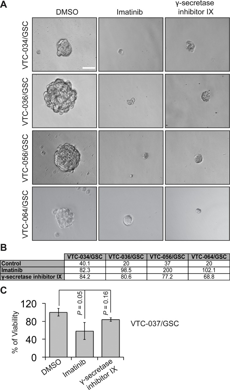
Figure 7. The effect of imatinib and γ-secretase inhibitor IX on patient-derived GSCs.
GSCs were plated at different cell densities and treated with vehicle DMSO, imatinib (10 μM), or γ-secretase inhibitor IX (20 μM). Cells were imaged using a 40 X lens of an inverted microscope (A). The sphere formation numbers were determined based on numbers of cells plated and the percentages of wells with no spheres (B). The responses of VTC-037/GSC to these drugs were determined using the MTS viability assay (C). Scale bar is 10 μm. Error bars represent standard deviations from three independent experiments.

