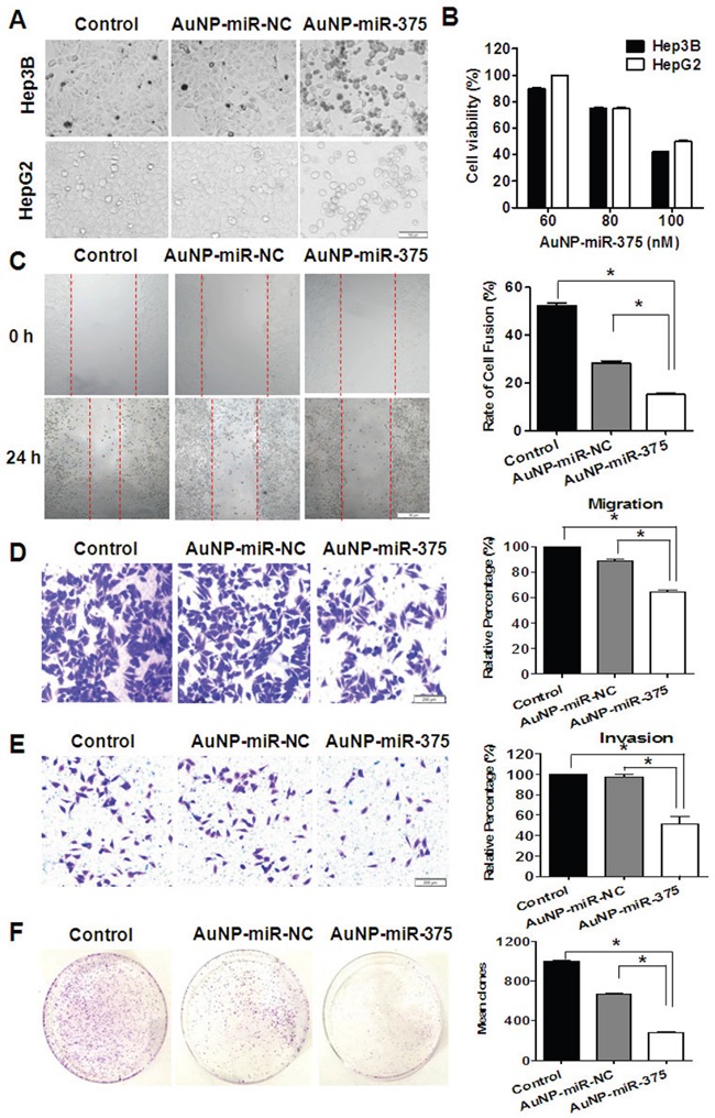
Figure 3. Anti-tumor activities of AuNP-miR-375 in hepatoma cells.

A. Morphology and cell viabilities were measured in HepG2 and Hep3B cells treated with AuNP-miR-375 (100 nM miR-375) or the controls for 48 h. The pictures were taken under an inverted light microscope (× 100). B. Viabilities of HepG2 and Hep3B cells treated with AuNP-miR-NC or AuNP-miR-375 for 48 h at different concentration of miR-375 as indicated. C. Wound-healing of Hep3B cells treated with AuNP-miR-NC or AuNP-miR-375 for 24 h. Quantitative results were calculated as: percent closure (%) = length of cell migration (mm) / width of wounds (mm) × 100%. Percent closure of control group was standardized as 100%. D. Transwell assay of Hep3B cells treated with AuNP-miR-NC or AuNP-miR-375. E. Matrigel invasion assay of Hep3B cells treated with AuNP-miR-NC or AuNP-miR-375. F. Colony formation assay of Hep3B cells. Hep3B cells were seeded in culture dishes for 2.5 × 10³ per well and allowed to grow for 15 days. All data are shown as mean ± SEM of 3 independent experiments. *: P < 0.05.
