
Figure 1. The expression of miRNAs derived from the Dlk1-Dio3 locus is upregulated in myostatin-deficient skeletal muscle.
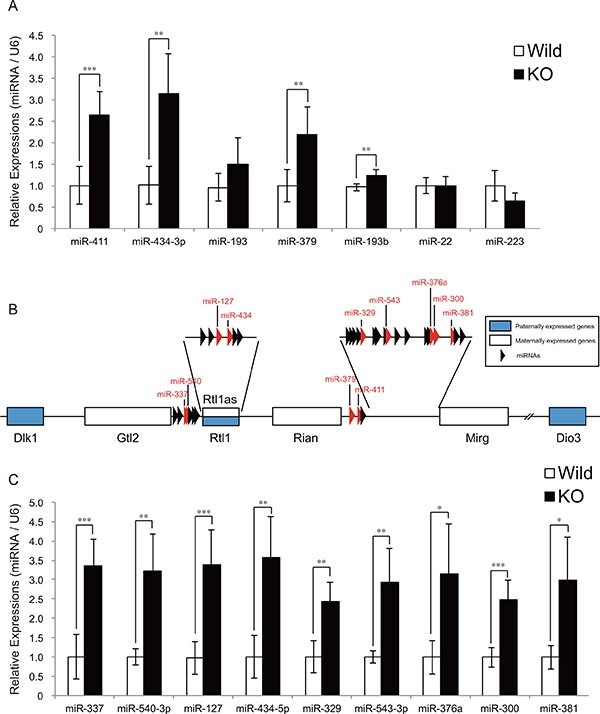
(A) A significant increase in miR-411, miR-434-3p, miR-379, and miR-193b expression in myostatin-deficient skeletal muscle at 13 weeks of age was determined by quantitative RT-PCR. (B) Schematic diagram of the Dlk1-Dio3 locus structure. This locus contains imprinting genes; Dlk1, Rtl1, and Dio3 (shown as blue boxes) are paternally expressed, whereas Gtl2, Rtl1as, Rian, Mirg (shown as white boxes), and miRNAs (arrowheads) are maternally expressed. Red arrowheads indicate the miRNAs whose expression was validated by qRT-PCR in Figure 1C. (C) Quantitative RT-PCR analysis shows a significant increase in the expression of miR-337, miR-540-3p, miR-127, miR-434-5p, miR-329, miR-543-3p, miR-376a, miR-300, and miR-381 in myostatin-deficient skeletal muscle at 13 weeks of age. The wild-type mice are the same age as the myostatin knockout mice. The results were normalized to U6 small RNA expression. Data are the mean ± SD (n = 5). *P < 0.05. **P < 0.01. ***P < 0.001.