Abstract
We herein report 3 cases of female patients with breast cancer who developed interstitial lung disease (ILD) during trastuzumab monotherapy in an adjuvant setting. Prior chemotherapy included 4 cycles of epirubicin and cyclophosphamide in patients 1 and 2, and 4 cycles of docetaxel, cyclophosphamide and trastuzumab in patient 3. Patient 1 presented with a cough and fever after the fourth cycle of trastuzumab. Patient 2 experienced rapid deterioration of oxygen saturation without subjective symptoms within 3 h of the first administration of trastuzumab. Patient 3 was unexpectedly diagnosed with organizing pneumonia in a scheduled computed tomography (CT) scan after the first course of trastuzumab. Based on clinical data, such as decreased PaO2 level, increased serum levels of KL-6 and/or lactate dehydrogenase, and findings on chest CT, these patients were diagnosed with drug-induced ILD. Considering the clinical course, trastuzumab was incriminated as the cause of ILD, particularly in patients 1 and 2. All 3 patients improved due to the timely diagnosis, discontinuation of trastuzumab and immediate administration of corticosteroid therapy. Although ILD is a rare adverse event associated with trastuzumab, it may cause rapid deterioration without preceding symptoms. Close observation and early diagnosis are required to avoid an unfavorable outcome.
Keywords: breast cancer, trastuzumab, interstitial lung disease, case reports
Introduction
Trastuzumab (Herceptin®) is a recombinant humanized immunoglobulin G1 monoclonal antibody against human epidermal growth factor receptor 2 (HER2), which is amplified in 15–30% of primary breast cancer patients (1,2). Incorporation of trastuzumab to adjuvant chemotherapy for early-stage HER2-positive breast cancer has been shown to reduce the risk of recurrence and to prolong survival (2–5). In metastatic patients, the combination of an anti-HER2 antibody, such as trastuzumab and/or pertuzumab, with a taxane has been established as the standard primary chemotherapy (6). It is thus widely accepted that trastuzumab is a backbone drug for the treatment of HER2-positive breast cancer. Trastuzumab is generally well-tolerated, but a proportion of patients develop serious complications, such as cardiac dysfunction and anaphylactic reactions. Although the association of interstitial lung disease (ILD) with trastuzumab has only rarely been reported, clinicians should be aware of the possibility of its serious clinical outcome (7). We herein report our recent experience with 3 breast cancer patients in whom trastuzumab monotherapy was complicated by ILD.
Case reports
Case 1
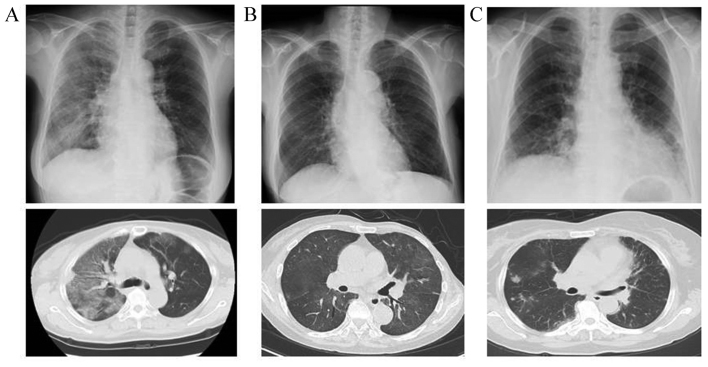
A 68-year-old female patient, with an Eastern Cooperative Oncology Group performance status (ECOG PS) score of 0, with no history of smoking or lung disease, underwent right breast quadrant resection. Pathological examination revealed invasive ductal carcinoma, scirrhous type, with negative sentinel node biopsy (T1N0M0). The cancer cells were estrogen receptor (ER)+, progesterone receptor (PR)+, and HER2+, with a Ki-67 index of 30%. The patient received 4 courses of 21-day-cycle adjuvant chemotherapy with epirubicin and cyclophosphamide, starting on the 46th postoperative day (POD), followed by triweekly trastuzumab. Anastrozole and radiotherapy to the right mammary area (total dose, 48 Gy) were started on the 185th and 192nd POD, respectively. The patient developed low-grade fever and cough on the 4th day of the 5th course of trastuzumab, corresponding to the day of 24/31 scheduled fraction of radiotherapy. Physical examination revealed coarse crackles on the whole of the right chest and the left upper chest. The laboratory tests revealed elevated levels of serum lactate dehydrogenase (LDH; 351 IU/l) and C-reactive protein (CRP; 15.3 mg/dl). The serum KL-6 level was within normal limits (247 IU/l). The PaO2 was 62 mmHg at room air (RA). The computed tomography (CT) scan revealed right lung-dominant patchy infiltration with surrounding ground-glass opacity (Fig. 1A). Infectious lung diseases were excluded by sputum cultures, Grocott's methenamine silver staining and polymerase chain reaction analysis of tuberculosis. The blood cultures and circulating galactomannan antigen tests were also negative. The patient was diagnosed with interstitial pneumonitis and was treated with steroid semi-pulse therapy with concurrent administration of meropenem and sulfamethoxazole/trimethoprim. The symptoms subsided within 7 days of the treatment.
Figure 1.
Chest X-ray (top) and computed tomography scan (bottom) at the onset of interstitial lung disease in patients (A) 1, (B) 2 and (C) 3.
Case 2
A 77-year-old female patient, with an ECOG PS score of 1, with no history of smoking or lung disease underwent left modified radical mastectomy. Pathological examination revealed invasive ductal carcinoma, scirrhous type, with negative sentinel node biopsy (T4bN0M0). The cancer cells were ER−, PR− and HER2+, with a Ki-67 index of 60%. The patient received 4 courses of 21-day-cycle adjuvant chemotherapy with epirubicin and cyclophosphamide, starting on the 68th POD, which was followed by trastuzumab monotherapy. After 3 h of the first administration of trastuzumab, the SpO2 fell rapidly to 81% at RA. Physical examination revealed a low-grade fever and right lung-dominant coarse crackles. The laboratory tests revealed elevated levels of serum LDH (552 IU/l), CRP (1.3 mg/dl) and KL-6 (719 IU/l). The PaO2 was 48.8 mmHg at RA. A CT scan revealed diffuse pale ground-glass opacities in the lungs bilaterally (Fig. 1B). Based on the radiological findings, the patient was diagnosed with interstitial pneumonitis and was treated with 30 mg of prednisolone, resulting in normalization of SpO2 within 7 days.
Case 3
A 62-year-old female patient, with an ECOG PS score of 1, with no history of smoking or lung disease, underwent right modified radical mastectomy. The patient had a history of gallbladder cancer, for which she underwent curative surgery 2 years prior to the onset of breast cancer. Pathological examination revealed invasive ductal carcinoma, solid-tubular type, with negative sentinel node biopsy (T2N0M0). The cancer cells were ER−, PR− and HER2+, with a Ki-67 index of 30%. The patient received 4 courses of 21-day-cycle adjuvant chemotherapy with docetaxel, cyclophosphamide and trastuzumab, starting on the 43rd POD. Although she experienced transient flu-like symptoms after the third course of the chemotherapy, the 4 courses of chemotherapy were completed, followed by trastuzumab monotherapy. On the 6th day of the first course of trastuzumab, a CT scan scheduled for a follow-up of the gallbladder cancer coincidentally revealed multiple patchy consolidations and ill-defined nodules in the lungs bilaterally (Fig. 1C). Physical examination revealed fine crackles bilaterally in the precordial chest, with SpO2 96% at RA. The laboratory tests revealed elevated levels of serum LDH (293 IU/l) and KL-6 (911 IU/l). Bronchoscopic lung biopsy revealed inflamed bronchial mucosa infiltrated by lymphocytes and histiocytes. Infection was excluded by various microbial tests of sputum and bronchoalveolar lavage specimens. The patient was diagnosed with drug-induced organizing pneumonia and was treated with 30 mg of prednisolone. The serum levels of KL-6 decreased gradually over 8 months.
Discussion
Although trastuzumab is indispensable to the treatment of HER2-positive breast cancer, physicians scarcely encounter trastuzumab-associated ILD. Previous large-scale trials, namely the B-31 and N9831 trials, which confirmed the efficacy of trastuzumab combined with paclitaxel after completing 4 courses of chemotherapy with doxorubicin and cyclophosphamide, reported that interstitial pneumonitis or pulmonary infiltrate was observed in 0.46 and 0.61% of patients, respectively (3). Although those studies suggested that trastuzumab possibly induces ILD when combined with cytotoxic anticancer drugs, the incidence of trastuzumab monotherapy-associated ILD has not been elucidated. In the large-scale phase 3 HERA trial, which verified the efficacy of trastuzumab monotherapy in an adjuvant setting, trastuzumab was administered to 3,105 patients without reports of ILD (2,4). Thus, although trastuzumab monotherapy-associated ILD is a recognized entity, it is a rare complication.
We herein reported the cases of 3 patients with trastuzumab monotherapy-associated ILD. Patient 1 developed ILD following administration of 5 courses of trastuzumab. Since breast radiotherapy was concurrently administered at the onset of ILD, the possibility of radiation pneumonitis cannot be excluded. However, a CT scan revealed a diffuse alveolar damage (DAD) pattern, spreading beyond the irradiated area. Furthermore, the onset of ILD was within 1 month of radiotherapy initiation, which is different from the typical clinical course of radiation pneumonitis. We thus considered trastuzumab to be the most likely cause of ILD in this patient. Patient 2 developed ILD 3 h after the first administration of trastuzumab, which was scheduled 3 weeks after the last cytotoxic chemotherapy with epirubicin and cyclophosphamide. Although the clinical symptoms were suggestive of an infusion reaction, the patient was diagnosed with trastuzumab-associated pneumonitis based on the findings of a CT scan, which corresponded well with the hypersensitivity pneumonitis (HP) pattern. Patient 3 developed ILD after administration of the first course of trastuzumab monotherapy following 4 cycles of combination chemotherapy with docetaxel, cyclophosphamide and trastuzumab. Although a scheduled CT scan as follow-up for gallbladder cancer coincidentally revealed ILD, it should be noted that this patient had experienced flu-like symptoms of unknown origin after the third course of the combination chemotherapy with docetaxel, cyclophosphamide and trastuzumab, and the LDH level was elevated 27 days prior to the emergence of ILD. Considering this clinical course, we hypothesized that the pre-existing drug-associated pulmonary inflammation became obvious and manifested as ILD after the initiation of trastuzumab monotherapy.
Thus far, several cases of ILD have been reported in association with combination chemotherapy with trastuzumab and cytotoxic drugs, such as paclitaxel and docetaxel (8,9). Those case reports mostly concluded that paclitaxel or docetaxel, rather than trastuzumab, was responsible for the development of ILD, as ILD is a well-known complication of taxanes. However, according to our literature survey, there have been 4 English-language case reports of trastuzumab monotherapy-associated ILD (10–13). Those cases and the 3 cases presented herein are summarized in Table I. The review of these cases suggests that ILD commonly manifests 3–4 months after trastuzumab treatment initiation. Furthermore, in all but 1 patient, trastuzumab monotherapy was initiated at an adjuvant setting after completing cytotoxic chemotherapy. It may be thus reasonable to hypothesize that the preceding cytotoxic chemotherapy in association with trastuzumab monotherapy may have affected the development of ILD. However, ILD appears to be a true complication of trastuzumab treatment, as at least some of the reported cases clearly suggest that trastuzumab was responsible for the development of ILD. Another important point is that radiological findings and clinical symptoms of trastuzumab-associated ILD appear to be variable. Indeed, our case series exhibited DAD, HP and OP pattern on CT. A previous review of antineoplastic drug-induced ILD estimated the incidence of trastuzumab-induced ILD to be as low as 0.4–0.6% (7). Despite its low incidence, clinicians should bear in mind that trastuzumab may induce ILD, which may manifest with variable non-specific patterns.
Table I.
Cases of trastuzumab monotherapy-induced ILD.
| Age/gender | Setting | Previous CRT | Duration after Tmab initiation | Pattern on CT | Treatment | Recovery | Refs. |
|---|---|---|---|---|---|---|---|
| 49/F | Adjuvant | AC, RT, D | 3 months | Focal (OP) | Steroid, antibiotics | Yes | (10) |
| 72/F | Palliative | None | 3 months | Diffuse (DAD) | Steroid | No | (11) |
| 65/F | Adjuvant | EC, PTX | 5 weeks | Diffuse (DAD) | Steroid, antibiotics | Yes (slow recovery) | (12) |
| 56/F | Palliative | DT | 19 weeks | Diffuse (CT: np) | Steroid | Yes | (13) |
| 68/F | Adjuvant | EC, RT | 3 months | Diffuse (DAD) | Steroid, antibiotics | Yes | Case 1 |
| 77/F | Adjuvant | EC | On first a administration | Diffuse (HP) | Steroid | Yes | Case 2 |
| 62/F | Adjuvant | DCT | 3 months | Focal (OP) | Steroid | Yes (slow recovery) | Case 3 |
ILD, interstitial lung disease; F, female; M, male; CRT, chemoradiotherapy; Tmab, trastuzumab; RT, radiotherapy; CT, computed tomography; np, not performed; D, docetaxel; AC, doxorubicin and cyclophosphamide; EC, epirubicin and cyclophosphamide; PTX, paclitaxel; DT, docetaxel and trastuzumab; DCT, docetaxel, cyclophosphamide and trastuzumab; OP, organizing pneumonia; DAD, diffuse alveolar damage; HP, hypersensitivity pneumonia.
Following diagnosis of drug-associated ILD, trastuzumab was discontinued in all 3 patients in the present study and steroid therapy was immediately initiated, resulting in recovery from drug-induced ILD. As shown in Table I, our literature survey suggests that trastuzumab-associated ILD is mostly reversible, resulting in a relatively favorable prognosis. Thus, early withdrawal of trastuzumab and immediate commencement of corticosteroid therapy are considered mandatory for a favorable outcome.
References
- 1.Slamon DJ, LeylandJones B, Shak S, Fuchs H, Paton V, Bajamonde A, Fleming T, Eiermann W, Wolter J, Pegram M, et al. Use of chemotherapy plus a monoclonal antibody against HER2 for metastatic breast cancer that overexpresses HER2. N Engl J Med. 2001;344:783–792. doi: 10.1056/NEJM200103153441101. [DOI] [PubMed] [Google Scholar]
- 2.Piccart-Gebhart MJ, Procter M, LeylandJones B, Goldhirsch A, Untch M, Smith I, Gianni L, Baselga J, Bell R, Jackisch C, et al. Trastuzumab after adjuvant chemotherapy in HER2-positive breast cancer. N Engl J Med. 2005;353:1659–1672. doi: 10.1056/NEJMoa052306. [DOI] [PubMed] [Google Scholar]
- 3.Romond EH, Perez EA, Bryant J, Suman VJ, Geyer CE, Jr, Davidson NE, Tan-Chiu E, Martino S, Paik S, Kaufman PA, et al. Trastuzumab plus adjuvant chemotherapy for operable HER2-positive breast cancer. N Engl J Med. 2005;353:1673–1684. doi: 10.1056/NEJMoa052122. [DOI] [PubMed] [Google Scholar]
- 4.Goldhirsch A, Gelber RD, PiccartGebhart MJ, de Azambuja E, Procter M, Suter TM, Jackisch C, Cameron D, Weber HA, Heinzmann D, et al. 2 years versus 1 year of adjuvant trastuzumab for HER2-positive breast cancer (HERA): An open-label, randomised controlled trial. Lancet. 2013;382:1021–1028. doi: 10.1016/S0140-6736(13)61094-6. [DOI] [PubMed] [Google Scholar]
- 5.Slamon D, Eiermann W, Robert N, Pienkowski T, Martin M, Press M, Mackey J, Glaspy J, Chan A, Pawlicki M, et al. Adjuvant trastuzumab in HER2-positive breast cancer. N Engl J Med. 2011;365:1273–1283. doi: 10.1056/NEJMoa0910383. [DOI] [PMC free article] [PubMed] [Google Scholar]
- 6.Baselga J, Cortés J, Kim SB, Im SA, Hegg R, Im YH, Roman L, Pedrini JL, Pienkowski T, Knott A, et al. Pertuzumab plus trastuzumab plus docetaxel for metastatic breast cancer. N Engl J Med. 2012;366:109–119. doi: 10.1056/NEJMoa1113216. [DOI] [PMC free article] [PubMed] [Google Scholar]
- 7.Vahid B, Marik PE. Pulmonary complications of novel antineoplastic agents for solid tumors. Chest. 2008;133:528–538. doi: 10.1378/chest.07-0851. [DOI] [PubMed] [Google Scholar]
- 8.Abulkhair O, El Melouk W. Delayed paclitaxel- trastuzumab-induced interstitial pneumonitis in breast cancer. Case Rep Oncol. 2011;4:186–191. doi: 10.1159/000326063. [DOI] [PMC free article] [PubMed] [Google Scholar]
- 9.Kuip E, Muller E. Fatal pneumonitis after treatment with docetaxel and trastuzumab. Neth J Med. 2009;67:237–239. [PubMed] [Google Scholar]
- 10.Radzikowska E, Szczepulska E, Chabowski M, Bestry I. Organising pneumonia caused by transtuzumab (Herceptin) therapy for breast cancer. Eur Respir J. 2003;21:552–555. doi: 10.1183/09031936.03.00035502. [DOI] [PubMed] [Google Scholar]
- 11.Vahid B, Mehrotra A. Trastuzumab (Herceptin)-associated lung injury. Respirology. 2006;11:655–658. doi: 10.1111/j.1440-1843.2006.00907.x. [DOI] [PubMed] [Google Scholar]
- 12.Bettini AC, Tondini C, Poletti P, Caremoli ER, Guerra U, Labianca R. A case of interstitial pneumonitis associated with Guillain-Barré syndrome during administration of adjuvant trastuzumab. Tumori. 2008;94:737–741. doi: 10.1177/030089160809400516. [DOI] [PubMed] [Google Scholar]
- 13.Pepels MJ, Boomars KA, van Kimmenade R, Hupperets PS. Life-threatening interstitial lung disease associated with trastuzumab: Case report. Breast Cancer Res Treat. 2009;113:609–612. doi: 10.1007/s10549-008-9966-8. [DOI] [PubMed] [Google Scholar]


