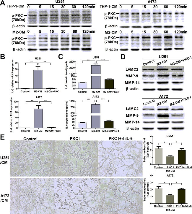
Figure 5. M2-enhanced IL-6 and VM in glioma cells via PKC pathway.

(A) Glioma cells were incubated with THP-1-CM or M2-CM for indicated time, the phosphorylation of PKC (p-PKC, Pan) was determined by Western blotting. (B, C) Glioma cells were incubated with DMEM medium, M2-CM or M2-CM containing Bisindolylmaleimide I (PKC I) for 24 h, IL-6 transcription (B) and concentration (C) in CM were determined by qRT-PCR and ELISA respectively. (D) As previously treatment, the protein levels of VM markers were then detected by Western blotting. (E) Representative images and quantification of tubule formation assay in glioma cells incubated in M2-CM, M2-CM containing Bisindolylmaleimide I or M2-CM containing Bisindolylmaleimide I with rhIL-6 for 24 h (×100). Each bar represents the mean ± SEM (n = 3). *P < 0.05; **P < 0.01; ***P < 0.001.
