
Figure 1. X-ray crystal structures of npcEBNA1.
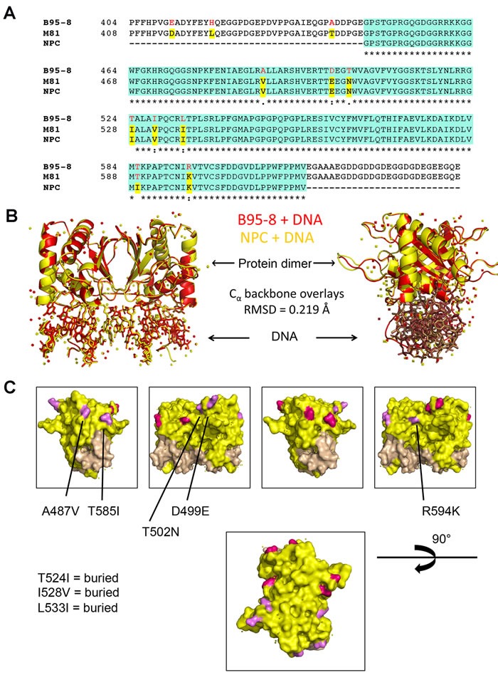
A. Alignment of primary amino acid sequence of EBNA1 from B95-8, M81, and NPC derivative. The DNA binding domain included in X-ray crystal structure is highlighted in green, polymorphisms are highlighted in red and yellow. B. X-ray crystal structure ribbon projection of NPC EBNA1 (yellow) superimposed on B95-8 EBNA1 (red) both bound to DNA, and with 90° rotation (right). C. Polymorphic amino acids that are surface exposed (A487V, T585I, T502N, D499E, R594K) are highlighted in magenta on one EBNA1 monomer and pink in the other EBNA1 monomer. Buried amino acids (T524I, I528V, L533I) are not seen on surface rendering.