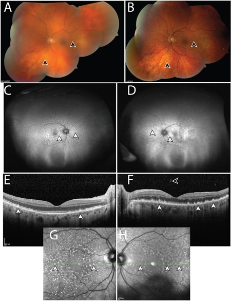
Figure 1. Manifestations of vitreoretinal lymphoma in Case 103.

A. Montaged fundus photo of the left eye with vitreous debris prior to intraocular liquid biopsy and vitrectomy. Lymphoma cells are suspended in the vitreous, resulting in a “hazy” view, which obscures anatomic details of the retina (arrowheads). B. Following intraocular liquid biopsy and vitrectomy, which did not detect malignant cells, the media of the left eye is clear and retinal details can be discerned, such as subretinal lipofuscin clumps, and sub-retinal pigment epithelium (RPE) deposits, which manifest in a yellow and dark stippled, leopard-like pattern (arrowheads). Ultra-wide field fundus autofluorescence of the right C. and left D. eye, shows stippled hyper-autofluorescence corresponding to the lymphomatous sub-RPE deposits (arrowheads). Optical coherence tomography of the right E. and left F. eye shows nodular hyperreflective lymphomatous lesions at the RPE level (arrowheads). Prior to biopsy of the left eye (F), lymphoma cells can be seen in the posterior vitreous. Insets G, H. represent near infrared reflectance imaging of the right (G) and left (H) eyes, which highlight the leopard-like pattern of the sub-RPE lymphomatous macular infiltrates. Green lines and arrowheads of insets (G, H) correspond to the cross sectional plane of the OCT images in (E) and (F). Similar to (A), autofluorescence (D), OCT (F), and near infrared reflectance imaging (H) in the left eye appear blurry compared to the right eye due to the presence of lymphoma cells in the vitreous. Except for (B), images were obtained following biopsy and vitrectomy in the right eye (C, E, G) but prior to these interventions in the left eye (A, D, F, H). During this time, visual acuity was within normal range.
