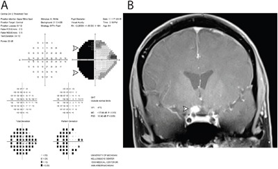
Figure 2. Functional and structural cause of vision loss in Case 103.

Two years following negative cytology results from intraocular liquid biopsies in each eye, the patient developed severe vision loss in the left eye. A. Humphrey 24-2 visual field testing report of the left eye revealed a dense temporal hemianopia, denoted by black area (arrowheads), which signified lack of sensitivity to light stimulus in the temporal half of visual field. B. Magnetic resonance imaging with gadolinium contrast revealed an enhancing lesion abutting the right optic chiasm and optic nerve. Subsequent cytological analysis of cerebrospinal fluid confirmed the diagnosis of primary CNS lymphoma, diffuse large-B cell subtype.
