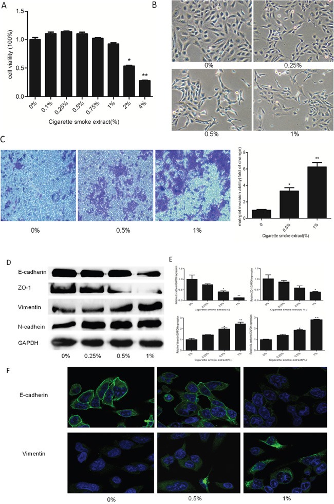
Figure 1. CSE induced EMT in SV-HUC-1 cells.

A. MTT assay showed cell viability decreased below 80% when cells were exposed to 2% or higher CSE concentrations in SV-HUC-1 cells. B. CSE induced morphological change from epithelial to spindle-like mesenchymal shape. SV-HUC-1 cells became longer and thinner, some of which generated slender tails. C. Transwell invasion assay revealed CS made a strong stimulative effect on the invasion capacity of SV-HUC-1 cells. The subsequent absorbance assay confirmed this change. D. CSE decreased the expression of epithelial markers E-cadherin and ZO-1, and increased expression of mesenchymal markers Vimentin and N-cadherin in SV-HUC-1 cells by Western blotting. E. CSE decreased the expression of E-cadherin and ZO-1 mRNAs, and enhanced the expression of Vimentin and N-cadherin mRNAs, detected by qRT-PCR. Data are expressed as mean ± SD. *p < 0.05, ** p < 0.01, compared with control group. F. Immunofluorescent staining also showed that CSE decreased E-cadherin protein expression and increased Vimentin expression in SV-HUC-1 cells.
