Abstract
Gram-negative, nonmotile bacteria that are catalase, oxidase, and urease positive are regularly isolated from the airways of horses with clinical signs of respiratory disease. On the basis of the findings by a polyphasic approach, we propose that these strains be classified as Nicoletella semolina gen. nov, sp. nov., a new member of the family Pasteurellaceae. N. semolina reduces nitrate to nitrite but is otherwise biochemically inert; this includes the lack of an ability to ferment glucose and other sugars. Growth is fastidious, and the isolates have a distinctive colony morphology, with the colonies being dry and waxy and looking like a semolina particle that can be moved around on an agar plate without losing their shape. DNA-DNA hybridization data and multilocus phylogenetic analysis, including 16S rRNA gene (rDNA), rpoB, and infB sequencing, clearly placed N. semolina as a new genus in the family Pasteurellaceae. In all the phylogenetic trees constructed, N. semolina is on a distinct branch displaying ∼5% 16S rDNA, ∼16% rpoB, and ∼20% infB sequence divergence from its nearest relative within the family Pasteurellaceae. High degrees of conservation of the 16S rDNA (99.8%), rpoB (99.6%), and infB (99.7%) sequences exist within the species, indicating that N. semolina isolates not only are phenotypically homogeneous but also are genetically homogeneous. The type strain of N. semolina is CCUG43639T (DSM16380T).
Bacteria belonging to the family Pasteurellaceae are commonly isolated from a number of different animals, including horses, and most are regarded as commensals or opportunistic pathogens. To date, nine different genera (Pasteurella, Actinobacillus, Haemophilus, Mannheimia, Lonepinella, Phocoenobacter, Histophilus, Gallibacterium, and Volucribacter) have been described within the family Pasteurellaceae. The genus Haemophilus is very heterogeneous and certainly needs taxonomic reorganization (13, 14). Species of this genus are isolated from various animals as well as humans, in which some can act as important pathogens. Mannheimia species are predominantly isolated from ruminants (3). Lonepinella koalarum is the only species described for the genus Lonepinella, and no host besides the koala bear has been reported (25). Similarly, Phocoenobacter uteri is the only species of the genus Phocoenobacter and was isolated from a harbor porpoise (12). Histophilus somni has been proposed to include the three species incertae sedis: “Haemophilus somnus,” “Haemophilus agni,” and “Histophilus ovis” (2). Christensen et al. (6) have described the genus Gallibacterium, which presently consists of one species and one genomospecies. Finally, Bisgaard Taxon 33 has been recently classified as Volucribacter (5). On the basis of present phylogenetic classification studies, additional genera in the family Pasteurellaceae are expected to be described in the future (15, 22, 23).
Except for the genera Pasteurella and Actinobacillus, no host-adapted member of the family Pasteurellaceae has been described for horses. “Pasteurella” caballi, misclassified as a member of the genus Pasteurella, is host associated and is sporadically found to be involved, along with other species, in respiratory infections of horses (8, 27). Bacteria belonging to the genus Actinobacillus are the most common isolates from the mucosal membranes of the oropharynx and respiratory tract in horses (26). Actinobacillus equuli has recently been divided into the two subspecies A. equuli subsp. equuli and A. equuli subsp. haemolyticus (7). Here we report on the characterization of a bacterium that has been repeatedly isolated from horses with airway disease. These isolates are phenotypically and phylogenetically distinct from the other members of the family Pasteurellaceae, so we propose the establishment of a new genus and species, Nicoletella semolina gen. nov., sp. nov.
MATERIALS AND METHODS
Bacterial strains and biochemical characterization.
All of the N. semolina strains used in this study are listed in Table 1. Strains used for DNA-DNA hybridization were Pasteurella multocida NCTC10322T, Actinobacillus lignieresii NCTC4189T, Actinobacillus capsulatus NCTC11408T, Actinobacillus equuli NCTC8529T, Actinobacillus ureae NCTC10220, Actinobacillus hominis SSI P575, Actinobacillus suis CCM5586T, Actinobacillus actinomycetemcomitans HIM946-2T, Pasteurella testudinis ATCC 33688T, Pasteurella pneumotropica ATCC 12555, Histophilus somni CCUG46774, and Haemophilus influenzae NCTC8143T. The strains included in the phylogenetic analyses are listed in the figures. All N. semolina strains originated from horses with clinical cases of airway disease. Strains either were isolated at the Institute of Veterinary Bacteriology from tracheal-bronchial washes of horses admitted to the Department of Equine Internal Medicine, University of Bern, from different regions within Switzerland or were received from the Culture Collection of the University of Göteborg (CCUG) (Table 1). Isolates were grown on chocolate agar with PolyViteX plates (bioMérieux Suisse S.A., Geneva, Switzerland) in an atmosphere of 5% CO2 for 24 to 48 h. Phenotypic characterization was done with API NH test strips (bioMérieux), according to the instructions of the supplier. As well, a limited number of isolates were also characterized by classical tube biochemical tests (23). For these tests, tubes were inoculated with a single colony, which was allowed to grow for 24 to 72 h, whereas in the API NH tests a large inoculum (turbidity equivalent to a 4 McFarland standard) of an overnight culture was used to inoculate the test strips for 2 h.
TABLE 1.
Strains used for the description of N. semolina isolated from diseased horses and assays performed
| Strain | Isolation yr | Origina | GenBank accession no. used for phylogenetic analysis
|
Assay performedc
|
||||
|---|---|---|---|---|---|---|---|---|
| 16S rDNA | rpoB | infB | API NH test | Biochemical characteristics | DNA-DNA hybridization | |||
| CCUG43639T | 1998 | CH | AY508816 | AY508861 | AY508835 | + | + | + |
| CCUG43640 | 1999 | CH | AY508817 | AY508862 | AY508836 | + | + | + |
| CCUG43646 | 2000 | CH | AY508818 | AY508863 | AY508837 | + | + | + |
| CCUG43638 | 1997 | CH | AY508819 | AY508864 | AY508838 | + | + | − |
| CCUG43641 | 1999 | CH | AY508820 | AY508865 | AY508839 | + | + | − |
| JF2465b | 2000 | CH | AY508821 | AY508866 | AY508840 | + | + | − |
| CCUG32135 | 1993 | S | AY508822 | AY508867 | AY508841 | + | − | − |
| CCUG39639 | 1998 | S | AY508823 | AY508868 | AY508842 | + | − | − |
| CCUG23468 | 1988 | S | AY508824 | AY508869 | + | − | − | |
| CCUG27342 | 1989 | S | AY508825 | AY508870 | + | − | − | |
| CCUG27497 | 1989 | S | AY508826 | AY508871 | + | − | − | |
| CCUG32179 | 1993 | S | AY508827 | AY508872 | + | − | − | |
| CCUG43643 | 1999 | CH | AY508828 | AY508873 | + | − | − | |
| CCUG43642 | 1999 | CH | AY508829 | AY508874 | + | − | − | |
| CCUG42978 | 1999 | S | AY508830 | AY508875 | + | − | − | |
| CCUG43647 | 2000 | CH | AY508831 | AY508876 | + | − | − | |
| CCUG43644 | 2000 | CH | AY508832 | AY508877 | + | − | − | |
| JF2408b | 1999 | CH | AY508833 | AY508878 | + | − | − | |
| CCUG43645 | 2000 | CH | AY508834 | AY508879 | + | − | − | |
CH, Switzerland; S, Sweden.
Strain number of the Institute of Veterinary Bacteriology, University of Bern.
+, assay was performed; −, assay was not performed.
DNA-DNA hybridization.
DNA-DNA hybridization was performed by the spectrophotometric method used by Mutters et al. (20). Renaturation rates of homologous and heterologous DNA solutions were determined with DNA at a concentration of 80 mg/ml in 2× SSC (1× SSC is 0.15 M NaCl plus 0.015 M sodium citrate) at 68°C.
Phylogenetic analyses.
Genomic DNA was isolated with a PUREGENE DNA extraction kit (Gentra Systems, Minneapolis, Minn.). The sequence of a 1.4-kb fragment of the 16S rRNA gene (rDNA) was determined as previously described by Kuhnert et al. (16, 17). A 560-bp fragment of the rpoB gene was amplified by PCR and directly sequenced by the method of Korczak et al. (15). Primers infB-L (ATGGGNCACGTTGACCACGGTAAAAC) and infB-R (CCGATACCACATTCCATACC) were designed for this study and were used for PCR amplification of a 1.3-kb fragment of the infB gene of all species except Phocoenobacter uteri, from which only a 0.5-kb fragment was obtained. The two PCR primers in combination with the internal primers infB-1 (CGTGAYGAGAARAAAGCACGTGAAG) and infB-2 (CTTCACGTGCTTTYTTCTCRTCACG) were used for further sequencing. Reactions were carried out in MicroAmp tubes (Applied Biosystems, Foster City, Calif.) on a GeneAmp 9600 thermal cycler (Applied Biosystems). For all three PCRs the cycling conditions were an initial 3 min of denaturation at 96°C, followed by 35 cycles of 30 s at 96°C, 30 s at 54°C, and 1 min at 72°C. A final extension step of 7 min at 72°C was included. The PCR products were purified with a High Pure PCR Purification kit (Roche Applied Science, Rotkreuz, Switzerland) prior to sequencing with a BigDye terminator cycle sequencing kit (Applied Biosystems). After purification of the sequencing products by ethanol precipitation, the samples were run on an ABI 3100 genetic analyzer (Applied Biosystems). The sequences of both strands were determined and edited by using Sequencher software (GeneCodes, Ann Arbor, Mich.). Phylogenetic relationships and trees were established with Bionumerics software (version 3.0; Applied Maths, Kortrijk, Belgium).
Nucleotide accession numbers.
The GenBank accession numbers for the 16S rDNA, rpoB, and infB sequences of the N. semolina strains determined in this study are listed in Table 1. The GenBank accession numbers for the infB sequences of the other species generated in this study are as follows: AY508843 for Gallibacterium anatis CCUG15563T, AY508844 for Pasteurella multocida subsp. multocida CCUG17976T, AY508845 for Volucribacter psittacicida JEO101, AY508846 for Lonepinella koalarum ATCC 700131T, AY508847 for Mannheimia haemolytica NCTC9380T, AY508848 for Histophilus somni HS8025T, AY508849 for Phocoenobacter uteri NCTC12872T, AY508850 for Actinobacillus genomospecies 1 CCUG22229, AY508851 for Actinobacillus pleuropneumoniae S4074T, AY508852 for Actinobacillus lignieresii NCTC4189T, AY508853 for Actinobacillus genomospecies 2 CCUG15571, AY508854 for Actinobacillus arthritidis CCUG24862T, AY508855 for Actinobacillus capsulatus CCUG12396T, AY508856 for Actinobacillus hominis CCUG19800T, AY508857 for Actinobacillus suis ATCC 33415T, AY508858 for Actinobacillus equuli subsp. haemolyticus CCUG19799T, AY508859 for Actinobacillus equuli subsp. equuli ATCC 19392T, and AY508860 for Actinobacillus ureae CCUG2139T.
RESULTS AND DISCUSSION
Bacteria that are fastidious in growth were isolated from the airways of diseased horses of different ages, including foals. These animals presented mainly with chronic cough and in a few cases with pneumonia and/or nasal discharge. For primary isolation, strains originating from tracheal-bronchial washes were grown on chocolate agar plates at 37°C in an atmosphere of 5% CO2. After 24 to 48 h of incubation, colonies resembling semolina particles could be observed. The gram-negative bacterium was a nonmotile, pleomorphic rod. It was the sole isolate or the predominant isolate and could be found at titers as high as 105/ml. The colonies, which were waxy, could be moved around the agar plate without losing their shape. These bacteria were difficult to cultivate directly from clinical material on 5% sheep or 5% horse blood agar plates but could be subcultured on these media. The organism did not grow on MacConkey agar plates. The N. semolina colonies were gray, showed no hemolysis on sheep-blood agar plates, and were nonadherent. The strains examined in this study have been isolated over several years from different countries, thereby representing a temporally, geographically, and epidemiologically independent set of isolates.
All of the strains were tested with the API NH system, a rapid test routinely used in many diagnostic laboratories. With this system, all strains were positive for d-glucose, d-fructose, d-saccharose, and urease and negative for penicillinase, l-ornithine, lipase, proline arylamidase, gamma glutamyltransferase, and indole. Seventy-five percent of the strains were d-maltose positive, while 65% of the strains were beta-galactosidase negative and 90% were alkaline phosphatase negative.
A preliminary analysis of the 16S rDNA of a few isolates indicated that these strains belong to the family Pasteurellaceae. Six strains were therefore further extensively analyzed for their biochemical properties by using classical tube tests useful for the phenotypic identification and discrimination of isolates within the family Pasteurellaceae (23). All strains were positive for nitrate reduction, as well as oxidase, catalase, and urease activities. Beta-galactosidase (o-nitrophenyl-β-d-galactopyranoside) and alkaline phosphatase reactions were variable but negative for most strains (for four and five of six strains, respectively). All strains were negative for arginine decarboxylase, lysine decarboxylase, and ornithine decarboxylase and for growth on citrate, adonitol, indole, H2S, and gelatinase. No acid was produced from any of the sugars tested, including l-sorbose, d-glucose, d-galactose, d-mannose, d-fructose, l-rhamnose, d-xylose, l-arabinose, d-sucrose, trehalose, maltose, d-lactose, raffinose, d-mannitol, d-sorbitol, dulcitol, and m-inositol; nor was acid produced from salicin and esculin. Starch was not hydrolyzed, and neither hemin (X-factor) nor NAD (V-factor) was required for growth. The biochemical reactions which can be used to differentiate N. semolina from the other genera of the family Pasteurellaceae are shown in Table 2.
TABLE 2.
Phenotypic criteria that allow differentiation of N. semolina from other genera of the family Pasteurellaceaea
| Test | Test result
|
|||||||||
|---|---|---|---|---|---|---|---|---|---|---|
| Nicoletella | Actinobacillus | Haemophilus | Pasteurella | Mannheimia | Histophilus | Lonepinella | Gallibacterium | Volucribacter | Phocoenobacter | |
| Catalase | + | + | + | + | + | − | − | + | v | − |
| Oxidase | + | + | + | + | + | + | − | + | v | + |
| Urease | + | + | v | v | − | − | − | − | − | − |
| Hemin (X factor) | − | − | + | − | − | − | − | − | − | − |
| NAD (V factor) | − | v | + | v | − | − | − | − | − | − |
| Indole | − | − | v | + | − | + | − | − | − | − |
| d-Galactose | − | + | + | + | + | − | + | + | + | − |
| d-Glucose | − | + | + | + | + | + | + | + | + | + |
Several differences in the sugar reactions (d-glucose, d-fructose, d-saccharose, and d-maltose) were observed between the commercial API NH system and the conventional tube format. These differences may be due to the fact that in the API NH system a large inoculum is used, and so no growth is required for the reaction. Therefore, the API NH test does not necessarily measure fermentation but, rather, measures only the acidification of sugars (e.g., indirectly by enzymatic sugar degradation). Therefore, the results obtained with the API NH test cannot be compared with those obtained by biochemical growth tests. For routine identification, the API NH system might give quick and reasonable results. However, for a scientifically sound discrimination of isolates within the Pasteurellaceae, the biochemical growth test results must be considered.
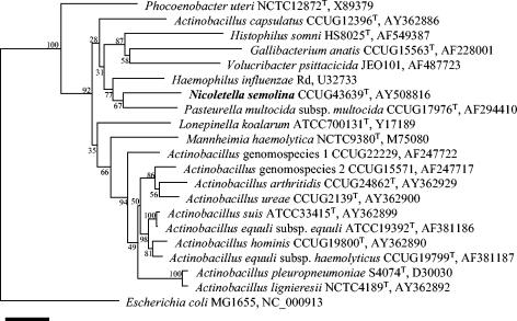
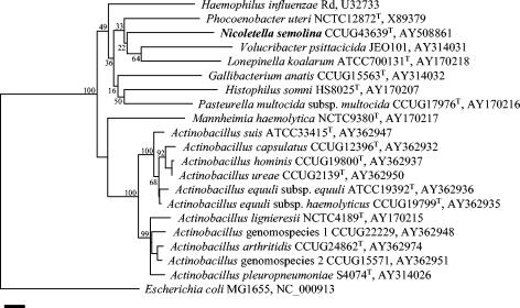
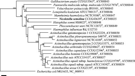
DNA-DNA hybridization studies were performed to investigate the relationship of N. semolina to other genera within the family Pasteurellaceae. Three strains of N. semolina, including the type strain, were hybridized against each other and showed binding values greater than 97%. As these were significant levels of DNA hybridization above the species level of 85% described for Pasteurellaceae (20), further hybridizations were done with only one representative of the group. Hybridization values for N. semolina with selected species of the Pasteurellaceae were highest with members of the genus Actinobacillus (from 54% with A. lignieresii to 66% with A. capsulatus). The genus Actinobacillus sensu stricto presently comprises the species A. lignieresii, A. suis, A. equuli subsp. equuli, A. equuli subsp. haemolyticus, A. pleuropneumoniae, A. ureae, A. arthritidis, Actinobacillus genomospecies 1 and 2, A. hominis, and A. capsulatus (21, 24). The position of A. capsulatus within the genus is still disputed, mainly because of its phylogenetic position within the 16S rDNA-derived tree (Fig. 1), but recent observations and the results presented here (Fig. 2 and 3) confirm the original classification by Mutters et al. (21). In order to further investigate whether the new taxon is a true member of Actinobacillus sensu stricto, additional phylogenetic investigations with species of this genus were undertaken to resolve the taxonomic position of this new taxon.
FIG. 1.
Phylogenetic tree based on 16S rDNA sequences. The sequence of N. semolina was compared phylogenetically to those of the type species of the Pasteurellaceae as well as the members of Actinobacillus sensu stricto. E. coli was chosen as an outgroup. The tree was built with Bionumerics software (version 3.0) by using the Jukes-Cantor correction and neighbor joining for cluster analysis. The accession numbers of the sequences used are given. Bootstrap values for 500 simulated runs are given. The solid bar indicates 2% sequence divergence.
FIG. 2.
Phylogenetic tree based on rpoB gene sequences. The sequence of N. semolina was compared phylogenetically to those of the type species of the Pasteurellaceae as well as the members of Actinobacillus sensu stricto. E. coli was chosen as an outgroup. The tree was built with Bionumerics software (version 3.0) by using the Jukes-Cantor correction and neighbor joining for cluster analysis. The accession numbers of the sequences used are given. Bootstrap values for 500 simulated runs are given. The solid bar indicates 2% sequence divergence.
FIG. 3.
Phylogenetic tree based on infB gene sequences. The sequence of N. semolina was compared phylogenetically to those of the type species of the Pasteurellaceae as well as the members of Actinobacillus sensu stricto. E. coli was chosen as an outgroup. The tree was built with Bionumerics software (version 3.0) by using the Jukes-Cantor correction and neighbor joining for cluster analysis. The accession numbers of the sequences used are given. Bootstrap values for 500 simulated runs are given. The solid bar indicates 2% sequence divergence.
Phylogenetic analysis was carried out, including comparison of the sequences of 16S rDNA, the gene for the β subunit of the RNA polymerase (rpoB), and the gene for translation initiation factor 2 (infB). The usefulness of 16S rDNA sequences for the establishment of genetic relationships within the family Pasteurellaceae was previously shown by Dewhirst and coworkers (10, 11). The rpoB gene has also been successfully applied for the elaboration of phylogenetic relationships in several groups of bacteria, including the Pasteurellaceae (2, 9, 15, 18). The efficacy of infB sequences was shown for the Pasteurellaceae by the phylogenetic delineation of the genera Haemophilus and Actinobacillus (13, 22). 16S rDNA analysis of N. semolina clearly showed that phylogenetically it belongs to the family Pasteurellaceae. Figure 1 shows the 16S rDNA-based tree for N. semolina, the nine presently described genera of this family, and the species of Actinobacillus sensu stricto in relation to Escherichia coli. N. semolina is positioned within the family Pasteurellaceae, yet it forms a branch of its own. This also holds true when N. semolina is included in the full 16S rDNA phylogeny (15). In order to compare the 16S rDNA sequence of N. semolina with all known 16S rDNA sequences, a search of the GenBank database was carried out with the BLAST algorithm (1). None of the 16S rDNA entries gave a greater than 95% match to N. semolina. Analysis of the rpoB genes resulted in phylogenetic relationships similar and complementary to those obtained with 16S rDNA. The results of rpoB sequence analysis are presented in the phylogenetic tree in Fig. 2, which again shows that N. semolina is clearly distinct from the other phyla. This also held true when N. semolina was included in the full rpoB phylogeny, since comparison of the N. semolina rpoB sequence with those from the entire family showed matches not greater than 85% (15). Finally, analysis of infB sequence-based phylogeny (Fig. 3) also showed a separate branching of N. semolina. The similarities of the N. semolina infB sequence to those of other known representatives were not greater than 80%.
The DNA-DNA hybridization values observed for the N. semolina strains (∼60%) with members of the genus Actinobacillus sensu stricto would allow classification of this new taxon in the genus Actinobacillus (20). However, the phenotypic and phylogenetic characteristics presented here argue for the classification of these strains as a new genus. First, the distinctive culture morphology and the inability to ferment d-glucose and other carbohydrates is so unusual (23) that these strains were at first not recognized as belonging to the family Pasteurellaceae (M. Bisgaard and J. Nicolet, personal communication). Nevertheless, the phylogenetic analyses of the strains as well as the DNA-DNA hybridization results clearly show that they belong to the family Pasteurellaceae. Second, the tree topologies obtained with the three genes analyzed did not show cobranching of N. semolina with the genus Actinobacillus. All species presently belonging to Actinobacillus sensu stricto were included in the phylogenetic analyses to emphasize this fact. Third, the sequence divergence of the genes of N. semolina analyzed to those of other species of the Pasteurellaceae and especially to the genus Actinobacillus was high enough to support a new genus (15, 22). In the case of 16S rDNA, this divergence from members of Actinobacillus sensu stricto was more than 5%. For rpoB, the divergence from Actinobacillus sensu stricto was about 18%, which is above the threshold value of 12% (15). Finally, in the case of infB, the divergence between N. semolina and members of the genus Actinobacillus sensu stricto was about 20%, which is again higher than that of the threshold value for this genus, which is 15% (22). On the other hand, phylogenetic analysis of numerous strains isolated from clinically, geographically and epidemiologically unrelated cases (Table 1) showed a high degree of conservation of the species genome, as represented by the three genes analyzed (Fig. 1 to 3). The intraspecies variabilities were very low: less than 0.2% for 16S rDNA, less than 0.4% for the rpoB gene, and 0.3% for the infB gene. This genetic conservation is also confirmed by the high DNA binding values of more than 97% between the three N. semolina strains analyzed; within other species of the Pasteurellaceae this value can be as low as 85% (20). N. semolina is therefore a highly homogeneous genetic group for which easy molecular confirmation of the preliminary phenotypic identification is possible, especially since identification by conventional biochemical growth tests is difficult. Furthermore, no differences in the major biochemical reactions or phenotypic criteria used to separate N. semolina from the other genera of Pasteurellaceae were observed (Table 2). Finally, the taxonomy of Pasteurellaceae is under constant revision since it contains many misclassified species. Phylogenetic analysis indicates that the number of genera at present must certainly be increased in order to structure the family reasonably. Placement of N. semolina into the genus Actinobacillus now would certainly make a future reclassification necessary, as we have shown by our phylogenetic analyses using accepted marker genes for the Pasteurellaceae.
Isolation of N. semolina in our experience is not uncommon. It remains to be determined whether N. semolina is a new emerging species or one that simply was not recognized in routine laboratory diagnostics in the past. Moreover, it will be important to determine whether N. semolina is a primary pathogen, is part of the normal flora that can occasionally cause disease, or is merely coincidentally present when there is airway disease. A retrospective analysis of putative N. semolina strains (strains of unknown designation which fit the description) that might reside in several laboratories, together with a prospective study of the prevalence of N. semolina in horse populations from various geographical regions, would help to clarify this point. A PCR approach based on one of the phylogenetic markers analyzed in this study could be helpful for that purpose. Future research will certainly be necessary to investigate the virulence potential of this new species and the pathogenesis of the disease associated with it.
Description of Nicoletella gen. nov.
Nicoletella (Ni.co.le.te′lla. N.L. fem. n. Nicoletella, named in tribute to Jacques Nicolet, a Swiss microbiologist, for his contribution to research on Pasteurellaceae).
Nicoletella is a new genus in the family Pasteurellaceae.
The genus Nicoletella consists of gram-negative, nonmotile, pleomorphic rods. Isolates do not grow on MacConkey agar and do not require hemin or NAD. They reduce nitrate to nitrite and are catalase, oxidase, and urease positive. Otherwise, the members of the genus are biochemically inert. They are negative for arginine decarboxylase, lysine decarboxylase, ornithine decarboxylase, adonitol, indole, H2S, and gelatinase and do not grow on citrate. No acid is produced from l-sorbose, d-glucose, d-galactose, d-mannose, d-fructose, l-rhamnose, d-xylose, l-arabinose, d-sucrose, trehalose, maltose, d-lactose, raffinose, d-mannitol, d-sorbitol, dulcitol, m-inositol, salicin, or esculin. Starch is not hydrolyzed. The type species is Nicoletella semolina.
Description of Nicoletella semolina sp. nov.
Nicoletella semolina (se.mo.li′na. N.L. fem. n. semolina, indicating the typical semolina-like colony characteristic).
Cells are gram-negative, nonmotile, pleomorphic rods. The species is capnophilic, and optimal growth conditions are on chocolate agar in a 5% CO2 atmosphere, on which after 24 to 48 h incubation it forms typical colonies of 0.5 to 2 mm in diameter which can be moved around on the plate like a semolina particle without losing their shape. The colony morphology is so typical that laboratory personnel can easily make a preliminary identification of this species. This is therefore an important diagnostic marker. The colonies are gray, odorless, and nonhemolytic and show positive reactions for catalase, oxidase, and urease. The species is biochemically inert when physiological characteristics are tested during growth. The species is negative for arginine decarboxylase, lysine decarboxylase, and ornithine decarboxylase and for growth on citrate, adonitol, indole, H2S, and gelatinase. No acid is produced from l-sorbose, d-glucose, d-galactose, d-mannose, d-fructose, l-rhamnose, d-xylose, l-arabinose, d-sucrose, trehalose, maltose, d-lactose, raffinose, d-mannitol, d-sorbitol, dulcitol, m-inositol, salicin, or esculin. Starch is not hydrolyzed.
With API NH test strips, N. semolina is positive for urease and acidification of glucose, fructose, and saccharose. Maltose, beta-galactosidase, and alkaline phosphatase reactions are variable. Tests for penicillinase, ornithine decarboxylase, lipase, proline amidase, gamma glutamyltransferase and indole are negative. Strains are isolated from the trachea of horses. The type strain is CCUG43639T (DSM 16380T).
Acknowledgments
We thank André Burnens for valuable discussions; Yvonne Schlatter, Carol Suter, Sandra Zumwald, and Margrit Krawinkler for excellent technical help; and Janet MacInnes and Sarah Burr for critical reading of the manuscript.
This research was supported by a grant (grant 6041.1 KTS) from KTI and by the Research Fund of the Institute of Veterinary Bacteriology, Bern, Switzerland.
REFERENCES
- 1.Altschul, S. F., W. Gish, W. Miller, E. W. Myers, and D. J. Lipman. 1990. Basic logical alignment search tool. J. Mol. Biol. 215:403-410. [DOI] [PubMed] [Google Scholar]
- 2.Angen, O., P. Ahrens, P. Kuhnert, H. Christensen, and R. Mutters. 2003. Proposal of Histophilus somni gen. nov., sp. nov. for the three species incertae sedis ‘Haemophilus somnus,’ ‘Haemophilus agni’ and ‘Histophilus ovis.’ Int. J. Syst. Evol. Microbiol. 53:1449-1456. [DOI] [PubMed] [Google Scholar]
- 3.Angen, O., R. Mutters, D. A. Caugant, J. E. Olsen, and M. Bisgaard. 1999. Taxonomic relationships of the [Pasteurella] haemolytica complex as evaluated by DNA-DNA hybridizations and 16S rRNA sequencing with proposal of Mannheimia haemolytica gen. nov., comb. nov., Mannheimia granulomatis comb. nov., Mannheimia glucosida sp. nov., Mannheimia ruminalis sp. nov., and Mannheimia varigena sp. nov. Int. J. Syst. Bacteriol. 49:67-86. [DOI] [PubMed] [Google Scholar]
- 4.Christensen, H., and M. Bisgaard. 2004. Revised definition of Actinobacillus sensu stricto isolated from animals. A review with special emphasis on diagnosis. Vet. Microbiol. 99:13-30. [DOI] [PubMed] [Google Scholar]
- 5.Christensen, H., M. Bisgaard, B. Aalbaek, and J. E. Olsen. 2004. Reclassification of Bisgaard taxon 33 with proposal of Volucribacter psittacicida gen. nov., sp. nov. and Volucribacter amazonae sp. nov. as new members of Pasteurellaceae. Int. J. Syst. Evol. Microbiol. 54:813-818. [DOI] [PubMed] [Google Scholar]
- 6.Christensen, H., M. Bisgaard, A. M. Bojesen, R. Mutters, and J. E. Olsen. 2003. Genetic relationships among avian isolates classified as Pasteurella haemolytica, ‘Actinobacillus salpingitidis’ or Pasteurella anatis with proposal of Gallibacterium anatis gen. nov., comb. nov. and description of additional genomospecies within Gallibacterium gen. nov. Int. J. Syst. Evol. Microbiol. 53:275-287. [DOI] [PubMed] [Google Scholar]
- 7.Christensen, H., M. Bisgaard, and J. E. Olsen. 2002. Reclassification of equine isolates previously reported as Actinobacillus equuli, variants of A. equuli, A. suis or taxon 11 of Bisgaard and proposal of two subspecies of A. equuli: A. equuli subsp. equuli and A. equuli subsp. haemolyticus. Int. J. Syst. Evol. Microbiol. 52:1569-1576. [DOI] [PubMed] [Google Scholar]
- 8.Church, S., K. E. Harrigan, A. E. Irving, and M. M. Peel. 1998. Endocarditis caused by Pasteurella caballi in a horse. Aust. Vet. J. 76:528-530. [DOI] [PubMed] [Google Scholar]
- 9.Dahllof, I., H. Baillie, and S. Kjelleberg. 2000. rpoB-based microbial community analysis avoids limitations inherent in 16S rRNA gene intraspecies heterogeneity. Appl. Environ. Microbiol. 66:3376-3380. [DOI] [PMC free article] [PubMed] [Google Scholar]
- 10.Dewhirst, F. E., B. J. Paster, I. Olsen, and G. J. Fraser. 1992. Phylogeny of 54 representative strains of species in the family Pasteurellaceae as determined by comparison of 16S rRNA sequences. J. Bacteriol. 174:2002-2013. [DOI] [PMC free article] [PubMed] [Google Scholar]
- 11.Dewhirst, F. E., B. J. Paster, I. Olsen, and G. J. Fraser. 1993. Phylogeny of the Pasteurellaceae as determined by comparison of 16S ribosomal ribonucleic acid sequences. Zentbl. Bakteriol. Parasitenkd. Infektkrankh. Hyg. Abt. 1 Orig. 279:35-44. [DOI] [PubMed] [Google Scholar]
- 12.Foster, G., H. M. Ross, H. Malnick, A. Willems, R. A. Hutson, R. J. Reid, and M. D. Collins. 2000. Phocoenobacter uteri gen. nov., sp. nov., a new member of the family Pasteurellaceae Pohl (1979) 1981 isolated from a harbour porpoise (Phocoena phocoena). Int. J. Syst. Evol. Microbiol. 50:135-139. [DOI] [PubMed] [Google Scholar]
- 13.Hedegaard, J., H. Okkels, B. Bruun, M. Kilian, K. K. Mortensen, and N. Norskov-Lauritsen. 2001. Phylogeny of the genus Haemophilus as determined by comparison of partial infB sequences. Microbiology 147:2599-2609. [DOI] [PubMed] [Google Scholar]
- 14.Kilian, M. 2004. Genus Haemophilus Winslow, Broadhurst, Buchanan, Krumwiede, Rogers and Smith 1917, 561AL. In R. Garrity (ed.), Bergey's manual of systematic bacteriology. Springer, New York, N.Y.
- 15.Korczak, B., H. Christensen, S. Emler, J. Frey, and P. Kuhnert. 2004. Phylogeny of the family Pasteurellaceae based on rpoB sequences. Int. J. Syst. Evol. Microbiol. 54:1393-1399. [DOI] [PubMed] [Google Scholar]
- 16.Kuhnert, P., S. E. Capaul, J. Nicolet, and J. Frey. 1996. Phylogenetic positions of Clostridium chauvoei and Clostridium septicum based on 16S rRNA gene sequences. Int. J. Syst. Bacteriol. 46:1174-1176. [DOI] [PubMed] [Google Scholar]
- 17.Kuhnert, P., J. Frey, N. P. Lang, and L. Mayfield. 2002. A phylogenetic analysis of Prevotella nigrescens, Prevotella intermedia and Porphyromonas gingivalis field strains reveals a clear species clustering. Int. J. Syst. Evol. Microbiol. 52:1391-1395. [DOI] [PubMed] [Google Scholar]
- 18.Mollet, C., M. Drancourt, and D. Raoult. 1997. rpoB sequence analysis as a novel basis for bacterial identification. Mol. Microbiol. 26:1005-1011. [DOI] [PubMed] [Google Scholar]
- 19.Mutters, R., H. Christensen, and M. Bisgaard. 2003. Genus Pasteurella Trevisian 1887, 94AL, Nom. cons. Opin. 13, Jud. Comm. 1954. In R. Garrity (ed.), Bergey's manual of systematic bacteriology. Springer, New York, N.Y.
- 20.Mutters, R., P. Ihm, S. Pohl, W. Frederiksen, and W. Mannheim. 1985. Reclassification of the genus Pasteurella Trevisan 1887 on the basis of deoxyribonucleic acid homology, with proposals for the new species Pasteurella dagmatis, Pasteurella canis, Pasteurella stomatis, Pasteurella anatis, and Pastuerella langaa. Int. J. Syst. Bacteriol. 35:309-322. [Google Scholar]
- 21.Mutters, R., W. Mannheim, and M. Bisgaard. 1989. Taxonomy of the group, p. 3-34. In C. Adlam and J. M. Rutter (ed.), Pasteurella and pasteurellosis. Academic Press, London, United Kingdom.
- 22.Norskov-Lauritsen, N., H. Christensen, H. Okkels, M. Kilian, and B. Bruun. 2004. Delineation of the genus Actinobacillus by comparison of partial infB sequences. Int. J. Syst. Evol. Microbiol. 54:635-644. [DOI] [PubMed] [Google Scholar]
- 23.Olsen, I., F. E. Dewhirst, B. J. Paster, and H. J. Busse. 2004. Family Pasteurellaceae. In R. Garrity (ed.), Bergey's manual of systematic bacteriology. Springer, New York, N.Y.
- 24.Olsen, I., and K. Moller. 2004. Genus Actinobacillus Brumpt 1919, 849AL. In R. Garrity (ed.), Bergey's manual of systematic bacteriology. Springer, New York, N.Y.
- 25.Osawa, R., F. A. Rainey, T. Fujisawa, E. Lang, H. J. Busse, T. Walsh, and E. Stackebrandt. 1995. Lonepinella koalarum gen. nov., sp. nov., a new tannin-protein complex degrading bacterium. Syst. Appl. Microbiol. 18:368-373. [Google Scholar]
- 26.Rycroft, A. N., and L. H. Garside. 2000. Actinobacillus species and their role in animal disease. Vet. J. 159:18-36. [DOI] [PubMed] [Google Scholar]
- 27.Schlater, L. K., D. J. Brenner, A. G. Steigerwalt, C. W. Moss, M. A. Lambert, and R. A. Packer. 1989. Pasteurella caballi, a new species from equine clinical specimens. J. Clin. Microbiol. 27:2169-2174. [DOI] [PMC free article] [PubMed] [Google Scholar]


