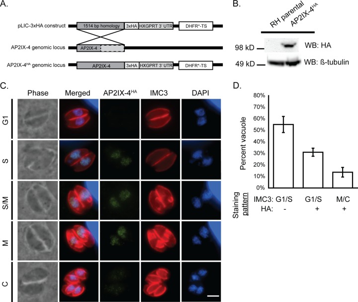
FIG 1 .
Characterization of AP2IX-4 protein expression. (A) Schematic showing the strategy used to generate parasites expressing AP2IX-4 tagged at the C terminus with 3xHA epitopes (AP2IX-4HA) in RHQ parasites. The AP2IX-4 genomic locus is aligned with the pLIC-3xHA construct used for endogenous tagging via single homologous recombination. The construct contains a 1,514-bp homology region and a DHFR*-TS drug selection cassette. UTR, untranscribed region. (B) Western blot (WB) of parental RHQ and AP2IX-4HA parasites probed with anti-HA antibody. Anti-β-tubulin was used to verify the presence of parasite protein. kD, kilodaltons. (C) IFAs performed on AP2IX-4HA tachyzoites using anti-HA (green). To monitor cell cycle phases, parasites were costained with anti-IMC3 (red) and DAPI (blue). G1, gap phase; S, synthesis phase; M, mitotic phase; C, cytokinesis. Scale bar, 3 µm. (D) Quantification of the proportion of parasite vacuoles (of 100) in the asynchronous population of AP2IX-4HA parasites exhibiting G1/S IMC3 staining with absence of HA signal (G1/S HA-), G1/S IMC3 staining with positive HA signal (G1/S HA+), or M/C staining with positive HA signal (M/C+). Error bars represent standard deviations of results from 3 independent experiments.

