Abstract
Proteins that self-assemble into regular shell-like polyhedra are useful, both in nature and in the laboratory, as molecular containers. Here we describe cryo-electron microscopy (EM) structures of two versatile encapsulation systems that exploit engineered electrostatic interactions for cargo loading. We show that increasing the number of negative charges on the lumenal surface of lumazine synthase, a protein that naturally assembles into a ∼1-MDa dodecahedron composed of 12 pentamers, induces stepwise expansion of the native protein shell, giving rise to thermostable ∼3-MDa and ∼6-MDa assemblies containing 180 and 360 subunits, respectively. Remarkably, these expanded particles assume unprecedented tetrahedrally and icosahedrally symmetric structures constructed entirely from pentameric units. Large keyhole-shaped pores in the shell, not present in the wild-type capsid, enable diffusion-limited encapsulation of complementarily charged guests. The structures of these supercharged assemblies demonstrate how programmed electrostatic effects can be effectively harnessed to tailor the architecture and properties of protein cages.
Self-assembling proteins that form capsid-like structures act as molecular containers for diverse cargoes. Here, the authors solve the cryo-EM structures of lumazine synthase shells, and show that supercharged mutants form expanded assemblies, indicating that electrostatics can be exploited to engineer cage architecture.
Hollow proteinaceous particles are widespread in nature. By providing a protective shell for molecular cargo, these nanostructures serve as multipurpose containers for the transport of viral payloads1, biomineralization2, protein folding3 and degradation4, enzyme encapsulation5,6 and even catalysis of short metabolic sequences7,8. Inspired by this precedent, protein cages are being engineered in the laboratory as customized nanoreactors9,10, delivery or display vehicles11 and artificial organelles12,13 for diverse applications in medicine, materials science and synthetic biology.
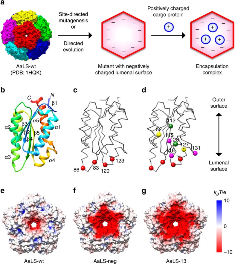
One highly versatile encapsulation system has been engineered from the capsid-forming enzyme lumazine synthase from the hyperthermophilic bacterium Aquifex aeolicus (Fig. 1a). Although the wild-type protein (AaLS-wt) adopts a flavodoxin-like αβ-fold14 (Fig. 1b) that bears no structural similarity to viral capsid proteins15,16,17,18, it spontaneously assembles into virus-like icosahedrally symmetric particles containing 60 identical subunits with a triangulation number19 T=1. In nature, these structures encapsulate riboflavin synthase, an enzyme that converts lumazine to riboflavin20. To bind unrelated guests, we mutated four residues that project into the lumen of the capsid to negatively charged glutamates. The resulting variant, called AaLS-neg (Fig. 1c,f), encapsulates proteins with complementary positive charges21. Loading capacity was subsequently improved roughly 10-fold by directed evolution, exploiting the ability of the cage to sequester a toxic protease and thereby protect the proteome of Escherichia coli22. The best variant, AaLS-13, possesses seven additional mutations that further increase the container's net negative charge (Fig. 1d,g) and enhance its ability to bind positively charged proteins both in vivo and in vitro22,23,24. The recent discovery that the encapsulation process is rapid enough to be limited by the rate of cargo diffusion highlights the unique properties of these supercharged protein assemblies25.
Figure 1. Design of the AaLS encapsulation system.
(a) Engineering AaLS cages to encapsulate cargo proteins based on electrostatic interactions. (b) A ribbon representation of the AaLS monomer. (c,d) Backbone traces of the AaLS-neg (c) and AaLS-13 (d) monomers in the same orientation as in b. Residues mutated in AaLS-neg are shown as red spheres (R83E, T86E, T120E and Q123E) (c), whereas the additional substitutes in AaLS-13 are shown as magenta (V115D, A118D and K131E), yellow (R52C, R127C) or green (D28G, T112S) spheres (d). (e–g) The lumenal surface of the AaLS-wt (e), AaLS-neg (f) and AaLS-13 (g) pentamers, showing electrostatic surface potentials assuming pH 8 and an ionic strength of 150 mM. For the variants, mutations were mapped onto the crystal structure of the wt pentamer.
Befitting their greater packaging capabilities, the negatively supercharged AaLS variants have substantially larger cage diameters than the native structure (AaLS-13, ∼40 nm; AaLS-neg, ∼30 nm; AaLS-wt, ∼16 nm)14,21,22. In analogy to viral proteins that can form icosahedral shells of varying size in response to external stimuli or cargo loading26,27, the structural transformations observed in these21,22 and other LS variants28 could conceivably reflect expansion of the starting T=1 particles to T=3 (180-mer), T=4 (240-mer) or even T=7 (420-mer) icosahedral structures29. However, the molecular mechanisms underlying these dramatic transitions and, more specifically, how protomer structure controls protein cage size and architecture are poorly understood.
Here, we elucidate the structures of the AaLS protein containers by native mass spectrometry (MS) and cryo-EM single-particle reconstruction techniques. Our results show that the expanded cages do not assemble according to well-established principles of quasi-equivalence19 but instead adopt unprecedented topologies that preserve some pentamer interfaces and leave other edges unsatisfied. Stable gaps in these shell structures rationalize why complementarily charged guests are encapsulated so efficiently.
Results
Native MS analysis
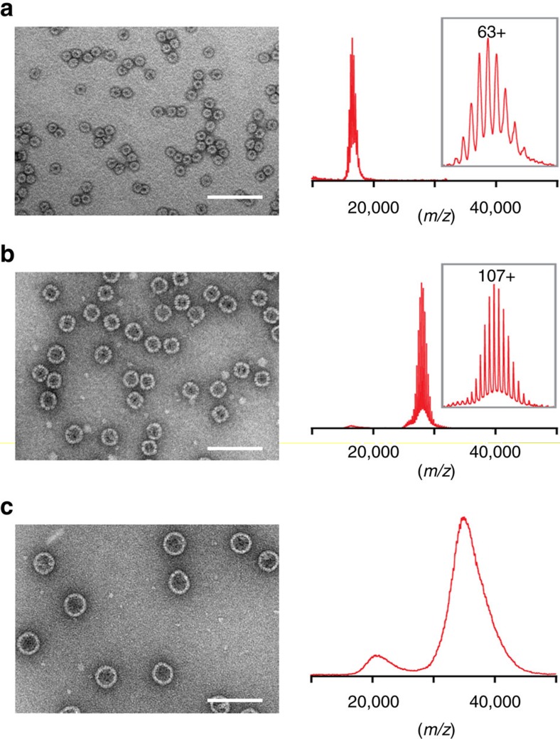
Native MS, which has provided accurate masses of viral capsids30, was employed to determine the subunit copy numbers of the AaLS capsids (Fig. 2). As expected, AaLS-wt gave a well-resolved charge state envelope corresponding to a 60-mer (measured mass of 1.06 MDa, theoretical mass of 1.05 MDa), consistent with the previously reported T=1 crystal structure14 (Fig. 2a; Supplementary Fig. 1a,b). For AaLS-neg, a similarly well-resolved spectrum was obtained that corresponds to a 180-mer (measured mass of 3.02 MDa, theoretical mass of 3.01 MDa) in agreement with the predicted icosohedral T=3 structure21 (Fig. 2b; Supplementary Fig. 1c,d). When the latter sample was pre-incubated in a low ionic strength buffer, however, only disassembled fragments corresponding to pentamers and smaller amounts of 10-mers and 15-mers were observed (Supplementary Fig. 1e,f); no dimeric, trimeric or hexameric building blocks, which would be expected for T=3 capsids31, were detected. Although no charge states could be resolved for AaLS-13, possibly because of structural heterogeneity, the MS data suggest that this cage contains between 240 and 420 subunits (Fig. 2c).
Figure 2. Size expansion of the AaLS variants.
(a–c) Negative-stain EM images and native mass spectra of AaLS-wt (a), AaLS-neg (b) and AaLS-13 (c). Scale bars, 100 nm. The most abundant charge states are annotated for AaLS-wt and AaLS-neg. The spectra clearly indicate that AaLS-wt is a 60-mer and AaLS-neg is a 180-mer. For AaLS-13, the oligomeric state was estimated to be between a 240-mer and a 420-mer extrapolating from the charging behaviour of the other AaLS variants.
Single-particle reconstruction
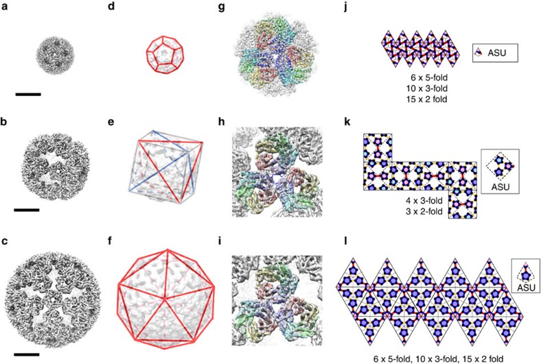
Encouraged by the apparent monodispersity of the AaLS-wt and AaLS-neg samples, cryo-EM single-particle reconstruction was performed to obtain detailed structural information32. The cryo-EM images of AaLS-wt were classified based on orientations of the particles in thin ice layers, and two-dimensional (2D) averages of each class were generated (Supplementary Fig. 2). The selected 2D class-averages were used to make an initial three-dimensional (3D) model by imposing icosahedral symmetry without any structural reference (Supplementary Fig. 2). This model was filtered to low-resolution and used as a reference for further 3D classification and refinement. The final EM density map of the AaLS-wt cage was calculated to 3.9 Å resolution from 3,268 particles (Fig. 3a), which allowed building and coordinate refinement of an atomic model (Fig. 3g; Table 1). The AaLS-wt cage is a 16-nm wide hollow dodecahedron consisting of 12 pentamers (Fig. 3d,j; Supplementary Fig. 5a). Our model agrees well with the T=1 crystal structure14 with a Cα root mean square deviation (RMSD) of 0.52 Å for all residues.
Figure 3. Cryo-EM structures of the AaLS variants.
(a–c) Cryo-EM density maps of AaLS-wt (a), AaLS-neg (b) and AaLS-13 (c) at 3.9, 5.4 and 5.2 Å resolution, respectively. Scale bars: 10 nm. (d–f) Dodecahedral, tetrahedral and icosahedral lattices matching the respective symmetries of AaLS-wt (d), AaLS-neg (e) and AaLS-13 (f). For AaLS-neg, two tetrahedral lattices (red and blue), embedded in a cube (grey), are depicted. (g–i) Close-up views of cryo-EM density maps of AaLS-wt (g), AaLS-neg (h) and AaLS-13 (i) around the three-fold rotational symmetry axes. Three pentamers from the refined models are shown as coloured ribbon diagrams. (j–l) 2D representation of the symmetry lattices for AaLS-wt (j), AaLS-neg (k) and AaLS-13 (l). Although the AaLS-neg structure is projected onto a flattened cube, note that it lacks the four-fold symmetry axes of a true cube because of the distinct pattern of pentamers on each face of the object and hence only possesses tetrahedral symmetry. The contact bars between two adjacent pentamers that have wt-like (pseudo) two-fold rotational symmetry are shown in red, while the other contact bars are shown in yellow. The asymmetric unit (ASU) for each structure is shown in the inset.
Table 1. Refinement and validation statistics for the atomic models of AaLS-wt, AaLS-neg and AaLS-13.
| Particle | AaLS-wt | AaLS-neg* | AaLS-13* |
|---|---|---|---|
| Data collection | |||
| No. of particles | 3,268 | 26,769 | 9,900 |
| Applied symmetry | icosahedral | tetrahedral | icosahedral |
| Pixel size (Å) | 1.39 | 1.39 | 1.39 |
| Defocus range (μm) | −1.2 to −3.4 | −1.2 to −3.4 | −1.2 to −3.4 |
| Voltage (kV) | 300 | 300 | 300 |
| Electron dose (e−/A2) | 25 | 25 | 25 |
| Reciprocal space data | |||
| Spacegroup | P1 | P1 | P1 |
| a=b=c (Å) | 252.00 | 420.00 | 448.00 |
| α=β=γ (°) | 90.0 | 90.0 | 90.0 |
| Refinement | |||
| Resolution range (Å) | 252.0–3.94 | 420.0–5.50 | 448.0–5.20 |
| Applied geometry weight (wxc) | 0.7 | 0.7 | 0.6 |
| No. reflections | 546,600 | 930,291 | 1,335,883 |
| R-factor | 0.250 | 0.215 | 0.248 |
| No. of protomers in ASU/particle | 1/60 | 15/180 | 6/360 |
| No. of residues | 9,240 | 28,980 | 57,960 |
| B-factor overall | 69.6 | 217.6 | 275.3 |
| R.m.s. deviations | |||
| Bond lengths (Å) | 0.009 | 0.008 | 0.008 |
| Bond angles (°) | 1.00 | 1.03 | 1.02 |
| Validation | |||
| Molprobity clashscore | 5.0 | 14.4 | 11.4 |
| Ramachandran plot | |||
| Favoured (%) | 94.7 | 91.9 | 90.2 |
| Allowed (%) | 3.3 | 6.5 | 7.0 |
| Outliers (%) | 0.0 | 1.6 | 2.8 |
*To stabilize the refinement, all atoms were used; however the final model does not include side chains because they are not resolved.
In the case of AaLS-neg, contrary to expectations for a conventional T=3 state21, imposing icosahedral symmetry during reconstruction did not result in interpretable density. By assuming tetrahedral symmetry, however, we obtained an initial reconstruction that was consistent with the distinct patterns of the 2D class-averages (Supplementary Fig. 3). This initial model was filtered to low-resolution and used for 3D reconstruction of the cage (Fig. 3b). The secondary structure elements of each subunit could be unambiguously assigned in the final EM density map refined to 5.4 Å resolution from 26,769 particles (Fig. 3h; Table 1). The 180 subunits of AaLS-neg, organized as 36 pentameric building blocks, form a novel 29-nm hollow tetrahedral cage with large, keyhole-shaped pores in the shell (Fig. 3e,k; Supplementary Fig. 5b). Such assemblies are unprecedented (Supplementary Table 1).
We next turned to the larger AaLS-13 assembly. Despite some sample heterogeneity, an initial model for AaLS-13 could be reconstructed from the 2D class-averages of selected particles by imposing icosahedral constraints without using a structural reference (Supplementary Fig. 4). The 3D classification, followed by refinement using 9,900 particles, resulted in an EM map resolved to 5.2 Å resolution (Fig. 3c). The characteristic αβ-fold of the LS monomer units was clearly evident (Fig. 3i; Table 1). Like its evolutionary parent, AaLS-13 has a highly porous cage structure constructed exclusively from pentameric building blocks, but, in contrast to AaLS-neg, it adopts icosahedral symmetry (Fig. 3f,l; Supplementary Fig. 5c). The 39-nm wide, ∼6-MDa large sphere is assembled from 72 pentamers (360 subunits), twice as many as AaLS-neg. Its internal volume (15,600 nm3) is 3.7 times larger than that of AaLS-neg (4,200 nm3) and 58 times larger than that of AaLS-wt (270 nm3), accounting for its enhanced loading capacity22.
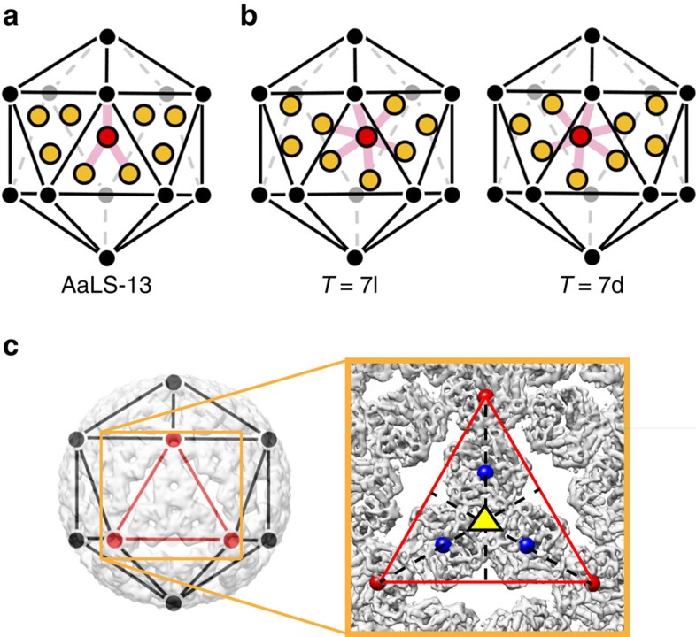
The AaLS-13 cage architecture can be best viewed as an extended dodecahedron, the dual counterpart of an icosahedron, with each face consisting of a central pentamer surrounded by five others (12 × 30-mer) (Supplementary Fig. 5d,e). Comparison of this structure with the icosahedral capsids of polyoma and papilloma viruses is instructive. The latter are also constructed from 72 pentameric building blocks, but the pentamers occupy both the pentavalent and hexavalent sites of a skewed T=7d icosahedral surface lattice33,34 (Fig. 4b). Due to differences in local environment, contacts between the capsomers at individual sites in the latter structures are not quasi-equivalent19 but mediated instead by the flexible tails of the shell proteins34. In contrast, the AaLS-13 assembly is not skewed (Fig. 4a,c), and its porous surface avoids offending contacts that could arise when a pentamer is packed inside a pseudohexagonal hole. Such topology has never been observed in nature (Supplementary Table 2). It is, however, reminiscent of the aperiodic pentagonal packings considered by Kepler in 1619 (ref. 35) and later by Penrose36.
Figure 4. AaLS-13 capsomer geometry.
(a,b) Positions of capsomers in AaLS-13 (a) compared with their locations in skewed T=7l and T=7d viral capsids (b). The twelve pentavalent vertices of the icosahedral lattice are shown as black and grey circles. For AaLS-13, pentameric capsomers, projected onto three triangular facets of the lattice, are shown as ochre or red circles; each interacts directly with only three other capsomers, as illustrated by the pink lines from the red unit. For the T=7l and 7d structures, the corresponding hexavalent sites are projected onto the same facets. In this case, the capsomer units have six neighbours each (shown by the pink lines from the red capsomer). (c) Overlay of an icosahedral lattice on the AaLS-13 structure. The cryo-EM density map viewed along one of the three-fold symmetry axes (yellow triangle) is shown magnified in the box on the right. Centroids of pentamers at five-fold symmetry axes are shown as red spheres. Centroids of pentamers projected onto an equilateral triangular facet of the icosahedral lattice are shown as blue spheres.
Stability
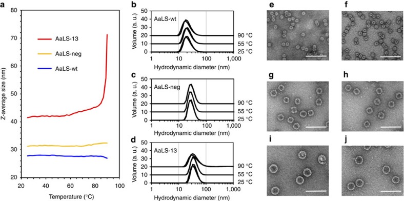
Wild-type AaLS is a remarkably thermostable protein (Tm=120 °C)14. Dynamic light scattering (DLS) measurements confirm that the AaLS-wt cage remains intact beyond 90 °C. Despite their highly porous nature, the mutant AaLS-neg and AaLS-13 cages are also quite stable (Fig. 5a–d). DLS measurements suggest that they do not disassemble below 80 °C, and negative-stain EM verified that the cage structures even survive heating for 5 min at 90 °C (Fig. 5e–j). Nevertheless, nanoindentation experiments using atomic force microscopy showed that the AaLS-13 cage is mechanically soft with a 20-fold smaller Young's modulus than AaLS-wt (ref. 37).
Figure 5. Thermal stability of the AaLS variants.
(a) DLS measurements with AaLS-wt (blue), AaLS-neg (ochre) and AaLS-13 (red). Z-Averages68 of the particles are plotted against the measured temperature (25–90 °C). The Z-averages of AaLS-neg (∼32 nm) and AaLS-13 (∼42 nm) correspond to the expected hydrodynamic diameters expected for 29- and 39-nm wide cages, respectively. However, the Z-average (∼28 nm) for AaLS-wt was significantly larger than the expected hydrodynamic diameter of a 16-nm wide cage, suggesting that a fraction of the particles aggregate in solution. (b–d) Volume weighted distribution of the hydrodynamic diameters determined for AaLS-wt (b), AaLS-neg (c) and AaLS-13 (d) at 25, 55 and 90 °C. The individual histograms are offset by 10 arbitrary volume units (a.u.) for clarity. Three independent measurements were performed and plotted for each condition. (e–j) Negative-stain EM images of AaLS-wt (e,f), AaLS-neg (g,h) and AaLS-wt (i,j). Each sample was deposited on the EM grid without heating (e,g,i) or immediately after heating for 5 min at 90 °C (f,h,j). Scale bars, 100 nm.
Discussion
Natural protein cages are usually organized based on symmetries present in cubic point groups. For instance, 12 and 24 copies of a single polypeptide can assemble into tetrahedrally and octahedrally symmetric objects2,38,39,40, respectively, whereas 60 polypeptides, or multiples thereof, form icosahedra5,20, as exemplified by viral capsids19,41. With few exceptions33,34,42,43,44, icosahedral structures contain 12 pentamers plus 10 × (T−1) hexamers, arranged to maintain quasi-equivalent inter-subunit interactions between individual polypeptides19.
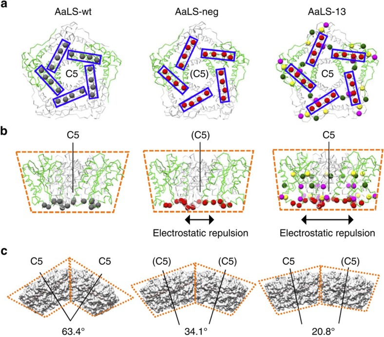
In the structures of the AaLS variants, the pentamer is the common and exclusive building block for all assemblies. While all pentamers in the wt cage are symmetrically equivalent, the AaLS-13 and AaLS-neg structures contain either two or three types of symmetrically non-equivalent pentamers, respectively (Fig. 3j–l; Supplementary Fig. 5). Superposition of AaLS-neg pentamers onto AaLS-wt pentamers revealed that the individual subunits of the engineered variant are slightly tilted outwards from the five-fold rotational symmetry axes (Cα RMSDs of 2.1–2.3 Å) (Supplementary Fig. 6d,f). This conformational change, which modifies the wedge-like shape of the pentamer (Fig. 6b), is likely the result of electrostatic repulsion between the anionic residues that were introduced on the lumenal surface (Fig. 6a,b and Supplementary Movie 1). Repositioning of the α4 helix (residues 120–131) (Supplementary Fig. 6a–c), which contains two of the designed mutations, further modifies the shape of the pentamer wedge. This secondary structural element appears to be relatively flexible, judging by its weak density and high B values, allowing it to adopt different conformations depending on the interaction partner. These effects are even more pronounced in AaLS-13 (Cα RMSDs of 2.8 Å) (Supplementary Fig. 6e,f), consistent with the increased number of anionic residues incorporated by directed evolution (Fig. 6a,b; Supplementary Movie 2). The A118D mutation, in particular, induces a substantial displacement of the α4 helix to avoid steric and electrostatic clashes.
Figure 6. Geometrical rearrangements triggered by tilted monomers.
(a,b) Representative pentamers of AaLS-wt, AaLS-neg and AaLS-13. Two of the five monomers for each structure are shown in green and the rest in grey. Views around the (pseudo) five-fold rotational symmetry axes (a) and the corresponding side views parallel to the axes (b) are shown. Residues 83, 86, 120 and 123, which were mutated to glutamates by design, are shown as red spheres and boxed. For AaLS-13, residues 28, 52, 112, 115, 118, 127 and 131, which were mutated during directed evolution, are shown as coloured spheres. Red and magenta spheres indicate the negatively charged residues that were introduced. The wedge-like shapes of the pentamers are schematically highlighted by dotted lines. (c) Representative orientation of two adjacent pentamers in AaLS-wt, AaLS-neg and AaLS-13, respectively. Angles between the adjacent local (pseudo) five-fold axes are shown.
How does the small conformational change in each pentamer trigger the observed global geometrical rearrangements? The modified pentamer wedges (Fig. 6b) would cause steric clashes if the cage maintained its original radius of curvature. To avoid such clashes, the angle between the local five-fold rotational symmetry axes of adjacent pentamers decreases, leading to formation of larger cages (Fig. 6c). The respective angles for AaLS-neg and AaLS-13 are in the range of 21.4–39.5° and 20.8–28.5°, significantly reduced from the 63.4° angle seen in AaLS-wt (Supplementary Fig. 7; Supplementary Movies 3,4).
The different angles found for the variants dictate the global structures and symmetries of the expanded cages. In the wt structure, two distinct inter-pentamer interfaces exist near the two- and three-fold rotational symmetry axes (Supplementary Fig. 8a–d). The three-fold interfaces located on the lumenal side of the cage are substantially reduced in size on increasing cage curvature (Supplementary Fig. 8e–g). In contrast, the two-fold interfaces near the outer surface of the cage are well maintained. Morph movies between AaLS-wt and the mutants show that these interfaces function as ‘hinges' between two pentamer wedges (Supplementary Movie 5). The position of one protomer is rotated ∼20° relative to its neighbour, changing the curvature of the cage while still maintaining contacts in the hinge region (Supplementary Fig. 9). Polymorphism in the cowpea chlorotic mosaic virus capsid, which can adopt different quasi-equivalent states, has been ascribed to a similar inter-subunit hinge movement45.
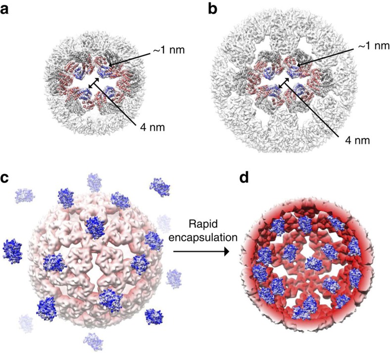
A remarkable feature of the negatively supercharged AaLS assemblies is that roughly 30% of the protomers (48/180 for AaLS-neg and 120/360 for AaLS-13) lack inter-pentamer interactions, leaving large pores in the shell wall. AaLS-neg and AaLS-13 possess 12 and 30 such openings, respectively, yet maintain high thermal stability. The solvent-exposed surfaces that define the edges of the keyhole-shaped slots do not show obvious architectural differences compared to their buried counterparts (Fig. 7a,b). At their centre, the openings are ∼4 nm wide and thus significantly larger than the channel located at the centre of each pentamer (<1 nm), allowing free passage across the shell for molecules with dimensions up to 4 nm. In combination with their supercharged interiors, this structural feature allows the AaLS mutants to function as ‘protein sponges' that absorb complementarily charged guest molecules at rates approaching the diffusion limit25 (Fig. 7c,d). Since AaLS-wt can also adopt expanded cage structures under certain conditions28, similar openings may mediate the release of enzymatically produced riboflavin, which is too large to pass through the <1 nm channels at the centre of the AaLS pentamers20.
Figure 7. Diffusion-limited protein cargo loading.
(a,b) The keyhole-shaped openings of AaLS-neg (a) and AaLS-13 (b). Each slot is surrounded by 16 protomers, 4 of which (shown in blue) lack the inter-pentamer interactions. (c,d) Scheme illustrating spontaneous encapsulation of supercharged GFP molecules by AaLS-13. A half-sliced AaLS-13 cage is depicted (d) to show association of the guest with the cage interior.
In nature, a single protein fold can be evolved to form multiple oligomeric states with different symmetries. The current study shows that such transformations can also be discovered by design and evolution in the laboratory. The novelty of the cage architectures studied here may serve as inspiration for ongoing efforts to create novel oligomeric protein assemblies. Completely artificial protein cages have been generated based on symmetries present in cubic point groups by designing building blocks with sterically complementary interfaces, either by site-directed mutagenesis46,47,48,49 or by using covalent linkages50,51. Formation of tetrahedrally and icosahedrally symmetric particles entirely from pentamers demonstrates that non-equivalent arrangements of individual building blocks may also be considered for the construction of isometric shells. The dramatic morphological changes observed in the supercharged AaLS variants additionally highlight the importance of mutations distant from the capsomer interface for determining overall cage architecture. By coupling such structural rearrangements in the individual building blocks to an external stimulus, such as a change in pH or ionic strength, it may even be possible to design structures that undergo controlled (dis)assembly.
The expanded repertoire of hierarchically ordered nanostructures formed by supercharged AaLS variants offers novel insights into the principles underlying the construction, function and evolution of protein assemblies. These highly plastic protein cages represent a versatile platform for creating tailored structures for a plethora of practical applications in nano- and medical science.
Methods
Preparation of AaLS variants
Plasmids encoding AaLS-wt, AaLS-neg, and AaLS-13 proteins were previously described21,22. Each protein was overexpressed with a C-terminal His6-tag in E. coli BL21-gold (DE3) cells and purified by Ni2+-NTA affinity chromatography (Qiagen) followed by size-exclusion chromatography using a HiPrep 16/60 Sephacryl S-400 HR column (GE Healthcare Life Sciences) with 20 mM Tris–HCl buffer (pH 7.8) containing 0.2 M NaCl and 1 mM EDTA according to published protocols29.
For native MS studies, AaLS-neg and AaLS-13 lacking a His6-tag were prepared. The respective genes were amplified from the plasmids encoding the corresponding His6-tagged variants by PCR with primers AaLS_fw (5′-AGA TAT ACA TAT GGA AAT CTA CGA AGG) and AaLS_noHis_rv (5′-GGT GGT GCT CGA GTC ATT ATT ATC GGA GAG ACT TGA ATA AGT TTG C). After digestion with NdeI and XhoI, each fragment was ligated into the original plasmid, which had been digested with the same restriction enzymes, to give pMG211_AaLS-neg_noHis and pMG211_AaLS-13_noHis. The proteins lacking the His6-tag were overexpressed in E. coli BL21-gold (DE3) cells, and the cell pellets from 0.4 l culture were resuspended in 15 ml of 50 mM sodium phosphate buffer (pH 8.0) containing 0.3 M NaCl, 1 mg ml−1 of lysozyme, 1 mg ml−1 of polymixin, EDTA-free protease inhibitor (complete protease inhibitor cocktail tablets, Roche), and a spatula tip of both DNase I and RNase A (Sigma Aldrich). After incubating at room temperature for one hour, the cells were disrupted by sonication and cleared by centrifugation. The crude lysates containing AaLS-neg were incubated at 85 °C for 20 min. For AaLS-13, 0.75 volumes of saturated aqueous ammonium sulphate were added to the lysates at room temperature. Following removal of the precipitate by centrifugation, the clear supernatants were buffer exchanged into 50 mM sodium phosphate buffer (pH 8.0) containing 0.2 M NaCl. The treated lysates were subjected to anion exchange chromatography using a MonoQ 10/100 GL column (GE Healthcare Life Sciences) preequilibrated with 50 mM sodium phosphate buffer (pH 8.0) containing 0.2 M NaCl and eluted by a linear gradient generated by increasing the NaCl concentration to 1 M. The protein fractions were buffer exchanged into 50 mM Tris–HCl buffer (pH 7.8) containing 1 M NaCl and concentrated until the monomer concentration exceeded 1 mM. After incubation over 7 days at room temperature, the protein samples were further purified by size-exclusion chromatography using a HiPrep 16/60 Sephacryl S-400 HR column with 50 mM Tris–HCl buffer (pH 8.0) containing 1 M NaCl as mobile phase. AaLS-13 samples were supplemented with 3 mM DTT to prevent formation of disulphide bonds.
Native MS
Purified AaLS variants were prepared for native MS by exchanging the buffer to 150 mM ammonium acetate (Sigma Aldrich), pH 7.8 through seven consecutive dilution and concentration steps using a 10-kDa molecular weight cutoff filter (Millipore). For native MS analysis, aliquots of the samples were diluted to a monomer concentration of 5 μM in 150 mM ammonium acetate, 25 mM triethylammonium acetate (Fluka), pH 7.8. For tandem MS experiments, aliquots of the samples were diluted in 150 mM ammonium acetate pH 7.8. For dissociation of AaLS-neg into smaller capsomers, samples were prepared in 10 mM ammonium acetate, pH 7.8. Experiments were performed on a quadrupole time-of-flight (qTOF) MS (MS Vision, Waters) modified for the analysis of high molecular weight ions52. AaLS-WT, AaLS-neg and AaLS-13 were introduced into the gas phase through the nano-electrospray interface using gold-coated borosilicate capillaries prepared in-house. Capillary voltages were set at ∼1,400 V in positive ion mode. Pressure in the xenon-filled collision cell was 1.8 × 10⁻² mbar. For native MS experiments, typical settings were 100 V sample cone voltage and 150–250 V collision voltage. For tandem MS experiments, typical settings were 50 V sample cone voltage and a collision voltage ramping from 50 to 300 V. The instrument was calibrated using caesium iodide clusters up to 30,000 m/z. Spectra were analysed using Masslynx 4.1 software (Waters). Masses were determined manually by minimization of the error over the charge state envelope from different charge state assignments.
Cryo-EM data collection
Purified samples (1.5–2.0 μl each) of AaLS-wt, AaLS-neg, and AaLS-13 cages (monomer concentration of 80, 80 and 300 μM, respectively) in 20 mM Tris–HCl buffer (pH 7.8) containing 0.2 M NaCl and 5 mM ethylenediaminetetraacetic acid (EDTA) were applied to holey carbon grids R2/2 (Quantifoil), which had been glow-discharged for 90 s using an Emitech K100X glow discharge system. The grids were blotted for 4 s twice at ambient temperature and 100% humidity and then plunged into liquid ethane using a Vitrobot mark one unit (FEI Company). Data were recorded semi-automatically using the EPU software on a Titan Krios transmission electron microscope (TEM) operated at 300 kV and equipped with a Falcon II direct electron detector (FEI Company). Images were acquired at −1.2 to −3.4 μm defocus at 100,720-fold magnification, resulting in a pixel size of 1.39 Å on the object scale. Beam-induced motion correction was further performed for images of AaLS-wt and AaLS-13. Specifically, the images were recorded as seven separate frames, comprising a total exposure of 25 electrons Å−2, which were subsequently aligned and summed using DOSEFGPU DRIFTCORR to obtain a final image53.
Cryo-EM data processing
Estimation of the contrast transfer function was performed using CTFFIND3 (ref. 54). Micrographs exhibiting poor power spectra based on the extent and regularity of the Thon rings were rejected. Particles were picked using EMAN 2.1 (ref. 55) or RELION 1.4 (ref. 56). 2D reference-free alignments of selected particles in 50 classes for AaLS-wt and AaLS-13 and 80 classes for AaLS-neg were performed by RELION 1.4. Particles that did not yield high-resolution class-averages were excluded from further refinement (Supplementary Figs 2–4 for more detailed workflows). Images of the selected 2D class-averages were used to reconstruct initial low-resolution models by imposing icosahedral symmetry for AaLS-wt and AaLS-13 or tetrahedral symmetry for AaLS-neg with the program e2initialmodel.py in EMAN 2.1. 3D classification was then performed with RELION 1.4 using the low-pass filtered initial models to 50 Å as references. Further 3D classification and refinement were performed using full-sized images (1.39 Å per pixel) and masks excluding the cage cavities.
Cryo-EM structural analysis
The cryo-EM maps and the molecular structures were analysed using UCSF Chimera 1.11 (ref. 57) and PyMOL (Version 1.7, Schrödinger LLC). Local resolutions of the EM maps were estimated using the ResMap program58. Electrostatic surface potentials were calculated for the crystal structure (PDB: 1HQK)14, for AaLS-wt, and mutation imposed crystal structure, for AaLS-neg and AaLS-13, at pH 8 and an ionic strength of 150 mM using PDB2PQR (refs 59, 60) and APBS (Adaptive Poisson-Boltzmann Solver)61 tools through UCSF Chimera. The default settings were used for the calculations except for the pH and ionic strength parameters. The graphics used for the figures and movies were generated using UCSF Chimera or PyMOL.
Atomic model building and refinement
For generation of a coordinate model that includes the entire AaLS-wt particle, one protomer of the corresponding crystal structure (PDB: 1HQK)14 was adjusted to the EM density using the program O (refs 62, 63) and symmetry expanded using the icosahedral operators from RELION 1.4 (ref. 56). The resulting model was subjected to phase-restrained reciprocal space refinement in PHENIX (ref. 64) (Table 1) against amplitudes and phases back-calculated from the 3.9 Å EM map, as described in ref. 65. During initial steps of coordinate and individual B-factor refinement, secondary structure and torsional non-crystallographic symmetry restraints were applied. The weighting of the coordinate geometry versus the map term was set to a value that resulted in a good overall model geometry during refinement (Table 1). For the final refinement round, one monomer was re-expanded, and the atomic model of the particle was minimized against the EM map imposing a strict 60-fold non-crystallographic symmetry. In the Fobs-Fcalc difference Fourier map, additional density can be identified close to the active site, which likely represents a reaction intermediate. However, as none of the ligands available from crystal structures solved in complex with substrate analogues66,67 convincingly explains the difference density and the resolution of the EM map does not allow modelling of a ligand at atomic detail de novo, we left this area of the map uninterpreted.
For the AaLS-neg and AaLS-13 mutants, the affected residues were exchanged in one of the 1HQK protomers, which then was expanded to 15 (AaLS-neg) or 6 (AaLS-13) positions to generate the asymmetric unit (ASU) of the particles. Areas of the ASU models located outside of the EM-density were manually readjusted in each monomer, which included a stretch around helix α4 (residues 117–134) and a neighbouring loop (residues 82–90). A few residues were added to the C-terminal α5 helix, which extends further towards the periphery of the particles. After tetrahedral 12-fold (AaLS-neg) or icosahedral 60-fold (AaLS-13) symmetry expansion, the full model was refined using a similar strategy as outlined above for AaLS-wt, except for B-factor refinement, which was performed in a ‘two groups per residue' mode (Table 1). Because of the limited resolution in the ∼5 Å range, in which only large bulky residues are visualized, the refined model was truncated to the backbone.
DLS
AaLS-wt, AaLS-neg and AaLS-13 purified by SEC were diluted in 50 mM Tris–HCl buffer (pH 7.8) containing 0.2 M NaCl and 5 mM EDTA to the final protein monomer concentration of 0.5 mg ml−1 (corresponding to 30 μM of AaLS monomer) before DLS analysis. The protein samples were measured at 25–90 °C with a 2 °C interval using a Zetasizer Nano ZS instrument (Malvern Instruments). Data analysis was performed using the Zetasizer software 7.11 (Malvern Instruments).
Negative-stain EM
AaLS-wt, AaLS-neg, and AaLS-13 purified by SEC were diluted in 20 mM Tris–HCl buffer (pH 7.8) containing 0.2 M NaCl and 1 mM EDTA to a final protein monomer concentration of 0.06–0.08 mg ml−1 (corresponding to 2–3 μM of AaLS monomer). The resulting samples were either left at room temperature or heated at 90 °C for 5 min in microcentrifuge tubes using Thermomixer Compact (Eppendorf). Immediately after heating or without heating, a 4-μl aliquot of each sample was deposited onto glow discharged copper grids with carbon support films (01814-F, Ted Pella). The grids were washed with the corresponding buffers and stained with 2% uranyl acetate before TEM analysis on a Morgagni 268 (FEI Company) microscope operated at 100 kV.
Data availability
The data that support the findings of this study are available from the corresponding author on reasonable request. The cryo-EM density maps have been deposited in the EM Data Bank with the accession codes EMD-3538 (AaLS-wt), EMD-3543 (AaLS-neg) and EMD-3544 (AaLS-13). Atomic coordinates of the corresponding models have been deposited in the Protein Data Bank with the accession codes 5MPP (AaLS-wt), 5MQ3 (AaLS-neg) and 5MQ7 (AaLS-13).
Additional information
How to cite this article: Sasaki, E. et al. Structure and assembly of scalable porous protein cages. Nat. Commun. 8, 14663 doi: 10.1038/ncomms14663 (2017).
Publisher's note: Springer Nature remains neutral with regard to jurisdictional claims in published maps and institutional affiliations.
Supplementary Material
Supplementary Figures and Supplementary Tables.
Morph movie between the AaLS-wt pentamer and the AaLS-neg pentamer. The mutated residues for AaLS-neg are shown as spheres, and the introduced negative charges (R83E, T86E, T120E, and Q123E) are indicated in red.
Morph movie between the AaLS-wt pentamer and the AaLS-13 pentamer. The mutated residues for AaLS-13 are shown as spheres, and the introduced negative charges from site-directed mutagenesis (R83E, T86E, T120E, and Q123E) and directed evolution (V115D, A118D, and K131E) are indicated in red and magenta, respectively. The other mutations from directed evolution are R52C and R127C (yellow), and D28G and T112S (green).
Morph movie between trimer of pentamers of AaLS-wt and AaLS-neg around 3-fold rotational symmetry axes. The mutated residues for AaLS-neg are colored as in Supplementary movie 1.
Morph movie between trimer of pentamers from AaLS-wt and AaLS-13 around the 3-fold rotational symmetry axes. The mutated residues for AaLS-13 are colored as in Supplementary movie 2.
Morph movie of the 2-fold contact between AaLS-wt and AaLS-13, showing the hinge motion. The 2-fold contact exits at the pentamer-pentamer interface of each structure
Acknowledgments
This work was supported by the European Research Council (Advanced ERC Grant ERC-dG-2012-321295 to D.H.), JSPS Postdoctoral Fellowships for Research Abroad (E.S.), the Stipendienfonds der Schweizerischen Chemischen Industrie (R.Z.), and the Netherlands Organization for Scientific Research (Proteins@Work project 184.032.201) and Fundamenteel Onderzoek der Materie (Projectruimte grant 12PR3303-2) to M.v.d.W. and A.J.R.H. The cryo-EM data were collected at the Scientific Center for Optical and Electron Microscopy (ScopeM, ETH Zürich) with excellent support from P. Tittmann. We thank R. Frey for his help with protein purification and T. Beck and A. Bach for technical assistance with the computational settings.
Footnotes
The authors declare no competing financial interests.
Author contributions All authors contributed to the experimental design. E.S. performed the cryo-EM experiments. E.S., D.B., M.L., N.B. and D.H. analysed the cryo-EM data. M.v.d.W. and A.J.R.H. carried out the native MS studies on samples prepared by R.Z. The manuscript was written by E.S. and D.H. and discussed and edited by the other authors.
References
- Flint J., Racaniello V. R., Rall G. F. & Skalka A. M. Principles of Virology ASM Press (2015). [Google Scholar]
- Liu X. & Theil E. C. Ferritins: dynamic management of biological iron and oxygen chemistry. Acc. Chem. Res. 38, 167–175 (2005). [DOI] [PubMed] [Google Scholar]
- Saibil H. R. Chaperone machines in action. Curr. Opin. Struct. Biol. 18, 35–42 (2008). [DOI] [PubMed] [Google Scholar]
- Kish-Trier E. & Hill C. P. Structural biology of the proteasome. Annu. Rev. Biophys. 42, 29–49 (2013). [DOI] [PMC free article] [PubMed] [Google Scholar]
- Sutter M. et al. Structural basis of enzyme encapsulation into a bacterial nanocompartment. Nat. Struct. Mol. Biol. 15, 939–947 (2008). [DOI] [PubMed] [Google Scholar]
- Bobik T. A., Lehman B. P. & Yeates T. O. Bacterial microcompartments: widespread prokaryotic organelles for isolation and optimization of metabolic pathways. Mol. Microbiol. 98, 193–207 (2015). [DOI] [PMC free article] [PubMed] [Google Scholar]
- Milne J. L. S. et al. Molecular architecture and mechanism of an icosahedral pyruvate dehydrogenase complex: a multifunctional catalytic machine. EMBO J. 21, 5587–5598 (2002). [DOI] [PMC free article] [PubMed] [Google Scholar]
- Maier T., Leibundgut M., Boehringer D. & Ban N. Structure and function of eukaryotic fatty acid synthases. Q. Rev. Biophys. 43, 373–422 (2010). [DOI] [PubMed] [Google Scholar]
- Bode S. A., Minten I. J., Nolte R. J. M. & Cornelissen J. J. L. M. Reactions inside nanoscale protein cages. Nanoscale 3, 2376–2389 (2011). [DOI] [PubMed] [Google Scholar]
- Jordan P. C. et al. Self-assembling biomolecular catalysts for hydrogen production. Nat. Chem. 8, 1–7 (2015). [DOI] [PubMed] [Google Scholar]
- Schoonen L. & van Hest J. C. M. Functionalization of protein-based nanocages for drug delivery applications. Nanoscale 6, 7124–7141 (2014). [DOI] [PubMed] [Google Scholar]
- Kim E. Y. & Tullman-Ercek D. Engineering nanoscale protein compartments for synthetic organelles. Curr. Opin. Biotechnol. 24, 627–632 (2013). [DOI] [PubMed] [Google Scholar]
- Frey R., Mantri S., Rocca M. & Hilvert D. Bottom-up construction of a primordial carboxysome mimic. J. Am. Chem. Soc. 138, 10072–10075 (2016). [DOI] [PubMed] [Google Scholar]
- Zhang X., Meining W., Fischer M., Bacher A. & Ladenstein R. X-ray structure analysis and crystallographic refinement of lumazine synthase from the hyperthermophile Aquifex aeolicus at 1.6 Å resolution: determinants of thermostability revealed from structural comparisons. J. Mol. Biol. 306, 1099–1114 (2001). [DOI] [PubMed] [Google Scholar]
- Reddy V. S. & Johnson J. E. Structure-derived insights into virus assembly. Adv. Virus Res. 64, 45–68 (2005). [DOI] [PubMed] [Google Scholar]
- Krupovic M. & Bamford D. H. Virus evolution: how far does the double beta-barrel viral lineage extend? Nat. Rev. Microbiol. 6, 941–948 (2008). [DOI] [PubMed] [Google Scholar]
- Abrescia N. G. A., Bamford D. H., Grimes J. M. & Stuart D. I. Structure unifies the viral universe. Annu. Rev. Biochem. 81, 795–822 (2012). [DOI] [PubMed] [Google Scholar]
- Suhanovsky M. M. & Teschke C. M. Nature's favorite building block: deciphering folding and capsid assembly of proteins with the HK97-fold. Virology 479–480, 487–497 (2015). [DOI] [PMC free article] [PubMed] [Google Scholar]
- Caspar D. L. D. & Klug A. Physical principles in the construction of regular viruses. Cold Spring Harb. Symp. Quant. Biol. 27, 1–24 (1962). [DOI] [PubMed] [Google Scholar]
- Ladenstein R., Fischer M. & Bacher A. The lumazine synthase/riboflavin synthase complex: shapes and functions of a highly variable enzyme system. FEBS J. 280, 2537–2563 (2013). [DOI] [PubMed] [Google Scholar]
- Seebeck F. P., Woycechowsky K. J., Zhuang W., Rabe J. P. & Hilvert D. A simple tagging system for protein encapsulation. J. Am. Chem. Soc. 128, 4516–4517 (2006). [DOI] [PubMed] [Google Scholar]
- Wörsdörfer B., Woycechowsky K. J. & Hilvert D. Directed evolution of a protein container. Science 331, 589–592 (2011). [DOI] [PubMed] [Google Scholar]
- Wörsdörfer B., Pianowski Z. & Hilvert D. Efficient in vitro encapsulation of protein cargo by an engineered protein container. J. Am. Chem. Soc. 134, 909–911 (2012). [DOI] [PubMed] [Google Scholar]
- Azuma Y., Zschoche R., Tinzl M. & Hilvert D. Quantitative packaging of active enzymes into a protein cage. Angew. Chem. Int. Ed. 55, 1531–1534 (2016). [DOI] [PubMed] [Google Scholar]
- Zschoche R. & Hilvert D. Diffusion-limited cargo loading of an engineered protein container. J. Am. Chem. Soc. 137, 16121–16132 (2015). [DOI] [PubMed] [Google Scholar]
- Cadena-Nava R. D. et al. Exploiting fluorescent polymers to probe the self-assembly of virus-like particles. J. Phys. Chem. B 115, 2386–2391 (2011). [DOI] [PubMed] [Google Scholar]
- He L., Porterfield Z., Van Der Schoot P., Zlotnick A. & Dragnea B. Hepatitis virus capsid polymorph stability depends on encapsulated cargo size. ACS Nano 7, 8447–8454 (2013). [DOI] [PMC free article] [PubMed] [Google Scholar]
- Zhang X. et al. Multiple assembly states of lumazine synthase: a model relating catalytic function and molecular assembly. J. Mol. Biol. 362, 753–770 (2006). [DOI] [PubMed] [Google Scholar]
- Sasaki E. & Hilvert D. Self-assembly of proteinaceous multishell structures mediated by a supercharged protein. J. Phys. Chem. B 120, 6089–6095 (2016). [DOI] [PubMed] [Google Scholar]
- Uetrecht C. et al. High-resolution mass spectrometry of viral assemblies: molecular composition and stability of dimorphic hepatitis B virus capsids. Proc. Natl Acad. Sci. USA 105, 9216–9220 (2008). [DOI] [PMC free article] [PubMed] [Google Scholar]
- Uetrecht C., Barbu I. M., Shoemaker G. K., van Duijn E. & Heck A. J. R. Interrogating viral capsid assembly with ion mobility-mass spectrometry. Nat. Chem. 3, 126–132 (2011). [DOI] [PubMed] [Google Scholar]
- Fernandez-Leiro R. & Scheres S. H. W. Unravelling biological macromolecules with cryo-electron microscopy. Nature 537, 339–346 (2016). [DOI] [PMC free article] [PubMed] [Google Scholar]
- Liddington R. C. et al. Structure of simian virus 40 at 3.8-Å resolution. Nature 354, 278–284 (1991). [DOI] [PubMed] [Google Scholar]
- Wolf M., Garcea R. L., Grigorieff N. & Harrison S. C. Subunit interactions in bovine papillomavirus. Proc. Natl Acad. Sci. USA 107, 6298–6303 (2010). [DOI] [PMC free article] [PubMed] [Google Scholar]
- Kepler J. Harmonices Mundi Libri V. (Linz, Austria (1619)) English translation: Kepler, J. The Harmony of the World. trans. Aiton, E. J., Duncan, A. M. & Field, J. V. (American Philosophical Society, 1997). [Google Scholar]
- Penrose R. The role of aesthetic in pure and applied mathematical research. Inst. Math. Appl. 10, 266–271 (1974). [Google Scholar]
- Heinze K. et al. Protein nanocontainers from nonviral origin: testing the mechanics of artificial and natural protein cages by AFM. J. Phys. Chem. B 120, 5945–5952 (2016). [DOI] [PubMed] [Google Scholar]
- Malet H. et al. Newly folded substrates inside the molecular cage of the HtrA chaperone DegQ. Nat. Struct. Mol. Biol. 19, 152–157 (2012). [DOI] [PMC free article] [PubMed] [Google Scholar]
- Borissenko L. & Groll M. Crystal structure of TET protease reveals complementary protein degradation pathways in prokaryotes. J. Mol. Biol. 346, 1207–1219 (2005). [DOI] [PubMed] [Google Scholar]
- van Montfort R. L., Basha E., Friedrich K. L., Slingsby C. & Vierling E. Crystal structure and assembly of a eukaryotic small heat shock protein. Nat. Struct. Biol. 8, 1025–1030 (2001). [DOI] [PubMed] [Google Scholar]
- Zandi R., Reguera D., Bruinsma R. F., Gelbart W. M. & Rudnick J. Origin of icosahedral symmetry in viruses. Proc. Natl Acad. Sci. USA 101, 15556–15560 (2004). [DOI] [PMC free article] [PubMed] [Google Scholar]
- Naitow H., Tang J., Canady M., Wickner R. B. & Johnson J. E. L-A virus at 3.4 Å resolution reveals particle architecture and mRNA decapping mechanism. Nat. Struct. Biol. 9, 725–728 (2002). [DOI] [PubMed] [Google Scholar]
- Kuhn R. J. et al. Structure of dengue virus: implications for flavivirus organization, maturation, and fusion. Cell 108, 717–725 (2002). [DOI] [PMC free article] [PubMed] [Google Scholar]
- Mukhopadhyay S., Kim B. S., Chipman P. R., Rossmann M. G. & Kuhn R. J. Structure of West Nile virus. Science 302, 248 (2003). [DOI] [PubMed] [Google Scholar]
- Tang J. et al. The role of subunit hinges and molecular ‘switches' in the control of viral capsid polymorphism. J. Struct. Biol. 154, 59–67 (2006). [DOI] [PubMed] [Google Scholar]
- King N. P. et al. Computational design of self-assembling protein nanomaterials with atomic level accuracy. Science 336, 1171–1174 (2012). [DOI] [PMC free article] [PubMed] [Google Scholar]
- King N. P. et al. Accurate design of co-assembling multi-component protein nanomaterials. Nature 510, 103–108 (2014). [DOI] [PMC free article] [PubMed] [Google Scholar]
- Hsia Y. et al. Design of a hyperstable 60-subunit protein icosahedron. Nature 535, 136–139 (2016). [DOI] [PMC free article] [PubMed] [Google Scholar]
- Bale J. B. et al. Accurate design of megadalton-scale two-component icosahedral protein complexes. Science 353, 389–394 (2016). [DOI] [PMC free article] [PubMed] [Google Scholar]
- Lai Y.-T., Cascio D. & Yeates T. O. Structure of a 16-nm cage designed by using protein oligomers. Science 336, 1129–1129 (2012). [DOI] [PubMed] [Google Scholar]
- Lai Y.-T. et al. Structure of a designed protein cage that self-assembles into a highly porous cube. Nat. Chem. 6, 1065–1071 (2014). [DOI] [PMC free article] [PubMed] [Google Scholar]
- Van den Heuvel R. H. H. et al. Improving the performance of a quadrupole time-of-flight instrument for macromolecualr mass spectrometry. Anal. Chem. 78, 7473–7483 (2006). [DOI] [PubMed] [Google Scholar]
- Li X. et al. Electron counting and beam-induced motion correction enable near-atomic-resolution single-particle cryo-EM. Nat. Methods 10, 584–590 (2013). [DOI] [PMC free article] [PubMed] [Google Scholar]
- Mindell J. A. & Grigorieff N. Accurate determination of local defocus and specimen tilt in electron microscopy. J. Struct. Biol. 142, 334–347 (2003). [DOI] [PubMed] [Google Scholar]
- Ludtke S. J., Baldwin P. R. & Chiu W. EMAN: semiautomated software for high-resolution single-particle reconstructions. J. Struct. Biol. 128, 82–97 (1999). [DOI] [PubMed] [Google Scholar]
- Scheres S. H. W. RELION: implementation of a Bayesian approach to cryo-EM structure determination. J. Struct. Biol. 180, 519–530 (2012). [DOI] [PMC free article] [PubMed] [Google Scholar]
- Pettersen E. F. et al. UCSF Chimera-A visualization system for exploratory research and analysis. J. Comput. Chem. 25, 1605–1612 (2004). [DOI] [PubMed] [Google Scholar]
- Kucukelbir A., Sigworth F. J. & Tagare H. D. Quantifying the local resolution of cryo-EM density maps. Nat. Methods 11, 63–65 (2014). [DOI] [PMC free article] [PubMed] [Google Scholar]
- Dolinsky T. J., Nielsen J. E., McCammon J. A. & Baker N. A. PDB2PQR: an automated pipeline for the setup of Poisson-Boltzmann electrostatics calculations. Nucleic Acids Res. 32, 665–667 (2004). [DOI] [PMC free article] [PubMed] [Google Scholar]
- Søndergaard C. R., Olsson M. H. M., Rostkowski M. & Jensen J. H. Improved treatment of ligands and coupling effects in empirical calculation and rationalization of pKa values. J. Chem. Theory Comput. 7, 2284–2295 (2011). [DOI] [PubMed] [Google Scholar]
- Baker N. A., Sept D., Joseph S., Holst M. J. & McCammon J. A. Electrostatics of nanosystems: application to microtubules and the ribosome. Proc. Natl Acad. Sci. USA 98, 10037–10041 (2001). [DOI] [PMC free article] [PubMed] [Google Scholar]
- Jones T. A., Zou J.-Y., Cowan S. W. & Kjeldgaard M. Improved methods for building protein models in electron density maps and the location of errors in these models. Acta Crystallogr. Sect. A 47, 110–119 (1991). [DOI] [PubMed] [Google Scholar]
- Jones T. A. Interactive electron-density map interpretation: from INTER to O. Acta Crystallogr. Sect. D Biol. Crystallogr. 60, 2115–2125 (2004). [DOI] [PubMed] [Google Scholar]
- Adams P. D. et al. PHENIX: a comprehensive Python-based system for macromolecular structure solution. Acta Crystallogr. Sect. D Biol. Crystallogr. 66, 213–221 (2010). [DOI] [PMC free article] [PubMed] [Google Scholar]
- Greber B. J. et al. The complete structure of the large subunit of the mammalian mitochondrial ribosome. Nature 515, 283–286 (2014). [DOI] [PubMed] [Google Scholar]
- Zhang X. et al. A structure-based model of the reaction catalyzed by lumazine synthase from Aquifex aeolicus. J. Mol. Biol. 328, 167–182 (2003). [DOI] [PubMed] [Google Scholar]
- Morgunova E. et al. Structural and thermodynamic insights into the binding mode of five novel inhibitors of lumazine synthase from Mycobacterium tuberculosis. FEBS J. 273, 4790–4804 (2006). [DOI] [PubMed] [Google Scholar]
- Koppel D. E. Analysis of macromolecular polydispersity in intensity correlation spectroscopy: the method of cumulants. J. Chem. Phys. 57, 4814 (1972). [Google Scholar]
Associated Data
This section collects any data citations, data availability statements, or supplementary materials included in this article.
Supplementary Materials
Supplementary Figures and Supplementary Tables.
Morph movie between the AaLS-wt pentamer and the AaLS-neg pentamer. The mutated residues for AaLS-neg are shown as spheres, and the introduced negative charges (R83E, T86E, T120E, and Q123E) are indicated in red.
Morph movie between the AaLS-wt pentamer and the AaLS-13 pentamer. The mutated residues for AaLS-13 are shown as spheres, and the introduced negative charges from site-directed mutagenesis (R83E, T86E, T120E, and Q123E) and directed evolution (V115D, A118D, and K131E) are indicated in red and magenta, respectively. The other mutations from directed evolution are R52C and R127C (yellow), and D28G and T112S (green).
Morph movie between trimer of pentamers of AaLS-wt and AaLS-neg around 3-fold rotational symmetry axes. The mutated residues for AaLS-neg are colored as in Supplementary movie 1.
Morph movie between trimer of pentamers from AaLS-wt and AaLS-13 around the 3-fold rotational symmetry axes. The mutated residues for AaLS-13 are colored as in Supplementary movie 2.
Morph movie of the 2-fold contact between AaLS-wt and AaLS-13, showing the hinge motion. The 2-fold contact exits at the pentamer-pentamer interface of each structure
Data Availability Statement
The data that support the findings of this study are available from the corresponding author on reasonable request. The cryo-EM density maps have been deposited in the EM Data Bank with the accession codes EMD-3538 (AaLS-wt), EMD-3543 (AaLS-neg) and EMD-3544 (AaLS-13). Atomic coordinates of the corresponding models have been deposited in the Protein Data Bank with the accession codes 5MPP (AaLS-wt), 5MQ3 (AaLS-neg) and 5MQ7 (AaLS-13).


