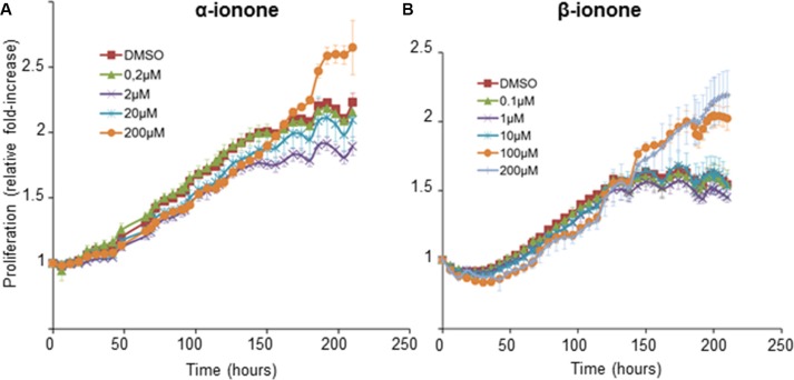
Figure 3. LNCaP cell growth induced by α-ionone or β-ionone.
LNCaP cells were seeded onto a collagen I gel in the presence of various concentrations of (A) α-ionone (0.2, 2, 20 or 200 μM) or (B) β-ionone (0.1, 1, 10, 100 or 200 μM), or of 0.1% DMSO (the amount of DMSO used to dilute odorants before adding them to the collagen gel or culture medium). Cell confluence was measured for 9 days and results are presented as an increase of proliferation during time (using proliferation=1 at t0). Bars indicate standard deviation (n = 3).

