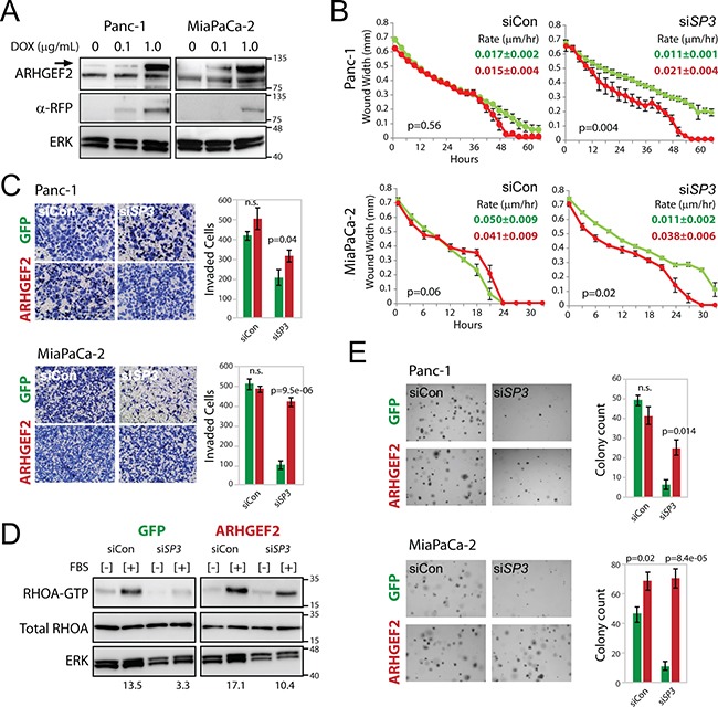
Figure 7. Enforced expression of ARHGEF2 rescues the SP3 knockdown phenotypes through restored activation of RHOA.

A. Western blot analysis of doxycycline induced cherry-ARHGEF2 (upper band, arrow) in Panc-1 and MiaPaCa-2 cells treated with doxycycline (DOX). Lysates were collected 24 hours post treatment and probed with antibodies against ARHGEF2 or red fluorescent protein (RFP). ERK served as the loading control. B. Wound width analysis of GFP expressing (green lines) and cherry-ARHGEF2 expressing (red lines) Panc-1 and MiaPaCa-2 cells transfected with control siRNA (siCon) or siRNA targeting SP3 (siSP3) monitored over the time course. Cells were grown in media supplemented with 0.1μg/mL doxycycline. Graphs were generated with the Essen IncuCyte ZOOM. P-values calculated from wound width analysis at 40 hour time point (Panc-1) and 18 hour time point (MiaPaCa-2). The rate constants for wound closure are indicated (μm/hr). C. Invasion of GFP and cherry-ARHGEF2 Panc-1 and MiaPaCa-2 cells through matrigel coated transwells following transfection with control siRNA (siCon) or siRNA targeting SP3 (siSP3). Cells were grown in media supplemented with 0.1μg/mL doxycycline. Bar graphs indicate the average number of invasive cells from three independent experiments of GFP expressing cells (green bars) and cherry-ARHGEF2 expressing cells (red bars). D. Western blot analysis of RHOA-GTP expression in Panc-1 cells expressing GFP or cherry-ARHGEF2 and transfected with siRNA control (siCon) or siRNA targeting SP3 (siSP3). Cells were grown in media supplemented with 0.1μg/mL doxycycline. 48 hours post siRNA transfection, lysates were collected following no stimulation [−] or 1 minute stimulation with FBS [+]. Lysates were incubated with Rhotekin-RHO binding domain protein beads, purified and subjected to western blot analysis. The ratio of RHOA-GTP/total RHOA is indicated. ERK served as the loading control. E. Representative images and quantification of GFP and cherry-ARHGEF2 Panc-1 and MiaPaCa-2 cells transfected with siRNA control (siCon) or siRNA targeting SP3 (siSP3). Cells were grown in media supplemented with 0.1μg/mL doxycycline prior to replating for colony assay. Cells were grown for 7-9 days in 0.3% agar to form colonies. Bar graphs show the average colony counts from three representative images of GFP expressing cells (green bars) and cherry-ARHGEF2 expressing cells (red bars).
