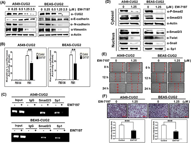
Figure 6. Treatment with EW-7197 inhibits the CUG2-induced EMT.

A. A549-CUG2 and BEAS-CUG2 cells were treated with EW-7197 at different doses (0.25, 0.5, 1.25 and 2.5 μM) for 24 h. Expression of CUG2, E-cadherin, N-cadherin, and vimentin was detected by immunoblotting. B. A549-CUG2 or BEAS-CUG2 cells were transfected with CUG2 promoter vectors (F961 and F961-94). At 48 h post-transfection, luciferase enzyme activities were measured in the transfected cell lysates. Transfection efficiency was normalized with the β-galactosidase reporter vector, pGK-β-gal. The assays were repeated in triplicate. The results shown are the average of triplicate wells. Error bars indicate SD. (**; p< 0.01) C. ChIP assays were performed with A549-CUG2 and BEAS-CUG2 cells. Chromatin fragments were pulled down with anti-Sp1, Smad2/3 antibodies or IgG as a control. Semi-quantitative PCRs were performed using specific CUG2 promoter primers. The assay was repeated twice. D. Expression of phospho-Smad2, Smad2/3, Snail and Twist in A549-CUG2 and BEAS-CUG2 cells treated with EW-7197 was detected by immunoblotting after cellular fractionations. Sp1 and actin were used loading controls for nuclear and cytosolic extracts, respectively. E. Cell migration was measured in A549-CUG2 and BEAS-CUG2 cells treated with EW-7197 by a wound healing assay. The assays were repeated twice. F. An invasion assay was performed with A549-CUG2 and BEAS-CUG2 cells treated with EW-7197. Scale bar indicates 100 μm. The assays were repeated twice. Each assay was performed in triplicate and error bars indicate SD. (***; p< 0.001).
