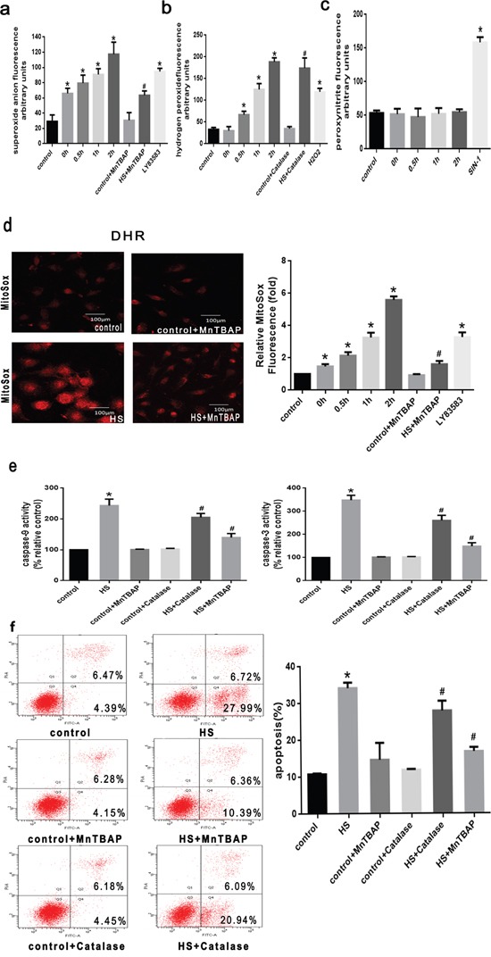
Figure 2. ROS involved in the mitochondrial apoptotic pathway is induced by intense heat stress in HUVEC cells.

a. O2.- production was measured with a commercial superoxide anion assay kit based on the oxidation of luminal, LY83583 (10μM) were used as positive control. b. H2O2 production was measured with peroxyfluor-6 acetoxymethyl ester (PF6-AM), H2O2 (25μM) were used as positive control. c. ONOO- was measured with luminol-amplified chemiluminescence, SIN-1(1mM) were used as positive control. d. Mitochondrial superoxide were labeled with MitoSOX™ Red, and Mitochondrial superoxide generation was labeled by laser scanning confocal microscopy. e-f. Cells were pretreated with MnTBAP (100μM) or Catalase (1000 U/μl) for 0.5h prior to heat stress (43°C) for 2h, and further incubated at 37°C for 6h. Enzymatic activity of caspase-9 and-3 was measured in cell lysates using the fluorogenic substrates Ac-LEHD-AFC and Ac-DEVD-AMC, respectively, and caspase activity was expressed relative to the control. Apoptosis induction was analyzed by flow cytometry using Annexin V-FITC/PI staining. Each value represents the mean ± SD of three separate experiments, *P < 0.05, relative to the control group (37°C), #P < 0.05, as compared to heat stress group (43°C).
