
Figure 5. Intense heat stress regulates Bcl-2 through superoxide in HUVEC cells.
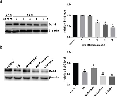
a. Cells were cultured at 43°C for 2h, then incubated at 37°C for different lengthes of time as indicated (0h, 1h, 3h, 6h or 9h). b. Cells were treated with 100μM MnTBAP or 1000 U/μl catalase for 0.5h prior to heat stress at 43°C for 2h, and further incubated at 37°C for 6h. 10μM LY83583 was used as positive control. Western blot analysis of Bcl-2 protein expression in HUVEC cells. Each value represents the mean ± SD of three separate experiments, *P < 0.05, compared to control group (37°C), #P < 0.05, compared to heat stress group (43°C).