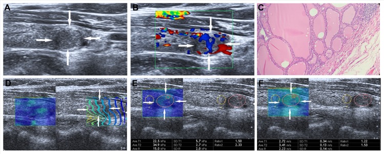
Figure 4. Images in a 55-year-old woman with nodular goiter.
(A) The nodule (arrows) is shown on conventional US, 10.1 mm × 6.8mm in size in the left lobe of the thyroid appears to have hypoechogenicity, well defined margin, regular shape, and without calcification. (B) The nodule (arrows) is shown on color-Doppler ultrasound, intra-nodular and slight peri-nodular blood flow. (C) (Haematoxylin-eosin stain, original magnification, ×200), Surgery-proven nodular thyroid goitre. (D) On the right, the nodule (arrows) shows regularly parallel contour lines on the shear wave propagation mode. (E) The E-mean, E-max of the nodule (arrows) expressed in kPa on SWS imagine are 22.5kPa and 34.9 kPa, respectively. (F) The E-mean, E-max of the nodule (arrows) expressed in m/s on SWS imagine are 2.72 m/s and 3.41 m/s respectively.

