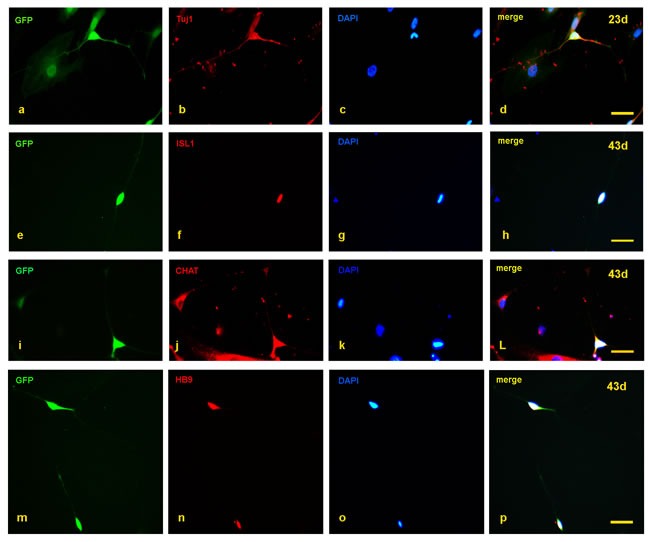
Figure 2. Detection of motor neuron markers with immunofluorescence staining.

a.-d. At day 23, the induced neurons expressed Tuj1. e.-p. At day 43, the induced neurons also expressed ISL1, CHAT, and HB9. Scale bars, 50 µm

a.-d. At day 23, the induced neurons expressed Tuj1. e.-p. At day 43, the induced neurons also expressed ISL1, CHAT, and HB9. Scale bars, 50 µm