Abstract
Objective: To present the rationale and detailed techniques for the application of exercises targeted to prevent anterior cruciate ligament (ACL) injury in high-risk female athletes.
Background: Female athletes have a 4- to 6-fold increased risk for ACL injury compared with their male counterparts playing at similar levels in the same sports. The increased ACL injury risk coupled with greater sports participation by young women over the last 30 years (9-fold increase in high school and 5-fold increase in collegiate sports) has generated public awareness and fueled several sex-related mechanistic and interventional investigations. These investigations provide the groundwork for the development of neuromuscular training aimed at targeting identified neuromuscular imbalances to decrease ACL injury risk.
Description: After the onset of puberty, female athletes may not have a neuromuscular spurt to match their similar, rapid increase in growth and development. The lack of a natural neuromuscular adaptation may facilitate the development of neuromuscular imbalances that increase the risk for ACL injury. Dynamic neuromuscular analysis training provides the methodologic approach for identifying high-risk individuals and the basis of using interventions targeted to their specific needs.
Clinical Advantages: Dynamic neuromuscular training applied to the high-risk population may decrease ACL injury risk and help more female athletes enjoy the benefits of sports participation without the long-term disabilities associated with injury.
Keywords: knee injury, injury-prevention training, neuromuscular training, dynamic knee valgus, neuromuscular imbalances
Anterior cruciate ligament (ACL) research has resulted in more than 2000 scientific articles outlining injury incidence, mechanism, surgical repair techniques, and rehabilitation of this important stabilizing knee ligament.1 However, despite the many scientific advances in the treatment of ACL injury, osteoarthritis occurs at a 10 times greater rate in individuals with ACL injury regardless of the treatment (conservative management versus surgical treatment).2 Epidemiologic research has demonstrated that female athletes have a 4- to 6-fold increased risk for ACL injury compared with their male counterparts playing at similar levels in the same sports.3,4 The increased ACL injury risk coupled with increased sports participation by young women over the last 30 years (9-fold increase in high school5 and 5-fold increase in collegiate sports6) has increased public awareness and fueled many sex-specific mechanistic and interventional investigations. This paradigm shift of isolated research focus away from treatment and rehabilitation is warranted, especially to injury mechanism and prevention, because an estimated 38 000 ACL injuries occur in young female athletes per year.7 At a cost per ACL injury of approximately $17 000,8 surgical and rehabilitative costs total approximately $646 000 000 annually in the United States. This is in addition to the traumatic effect to these individuals of potential loss of entire seasons of sports participation and possible scholarship funding, significantly lowered academic performance,9 long-term disability, and up to 105 times greater risk for radiographically diagnosed osteoarthritis in the future.10
Contrary to what is observed in adolescent athletes, a thorough analysis of the published literature demonstrates no evidence that a difference in ACL injury rates exists in prepubescent athletes.11–14 Knee injuries do occur in pediatric athletes, as evidenced by 63% of the sport-related injuries in children aged 6–12 years being classified as joint sprains, most of which occur at the knee.14 However, ACL sprains are more rare in prepubescent children than in adolescents, and ruptures do not present at significantly different rates in boys and girls before puberty.11–13 Although equal numbers of ligament sprains occur in girls and boys before adolescence, girls have higher rates immediately after their growth spurt and into maturity.15
The anthropometric measures of growth and development show very similar trends between the sexes, but male and female force-production capabilities diverge significantly during and after puberty. Men demonstrate a neuromuscular spurt, whereas women, on average, exhibit little change throughout puberty.16,17 The neuromuscular spurt is defined as increased power, strength, and coordination that occur with increasing chronologic age and maturation stage in adolescent boys.16,17 No similar correlations among height, weight, and neuromuscular performance have been demonstrated in pubescent girls. For example, vertical jump height (a measure of whole-body power) increases steadily in boys during puberty but not in girls.16–19 No sex differences in peak leg power were noted before age 14, but boys have significantly greater power after that age.20 A plateau in the peak power of girls occurred around 16 years of age.20 In the absence of appropriate neuromuscular adaptation, the musculoskeletal growth during puberty may increase neuromuscular imbalances. We define neuromuscular imbalances as muscle strength or activation patterns that lead to increased joint load. Female athletes may demonstrate one or more neuromuscular imbalances that increase lower extremity joint loads during sports activities.21,22 With ligament dominance, the neuromuscular and ligamentous control of the joint is unbalanced, as demonstrated by an inability to control dynamic knee valgus when landing and cutting.23 Quadriceps dominance relates to an imbalance between knee extensor and flexor strength, recruitment, and coordination.22 With leg dominance, the 2 lower extremities are unbalanced in strength and coordination.21,23 These developmental imbalances, if left unchecked, may continue through adolescence into maturity. Huston and Wojtys24 showed that college-aged elite female athletes, well past their developmental years, demonstrated neuromuscular recruitment and leg-strength imbalances when compared with male athletes and nonathletic controls.
Neuromuscular imbalances may be important contributors to ACL injury, which occurs under conditions of high dynamic loading of the knee joint, when active muscular restraints do not adequately compensate and dampen joint loads.25 Decreased neuromuscular control of the joint may stress the passive ligament structures, exceeding the failure strength of the ligament.26,27 High levels of neuromuscular control are necessary to create dynamic knee stability.26,28 Any neuromuscular imbalances that limit the effectiveness of the active muscular control system in working synergistically with the passive joint restraints to create dynamic knee stability may increase the risk for an ACL injury. Identifying these imbalances may offer the greatest potential for interventional development and application in high-risk populations.29 Our purpose is to present the theory and rationale of methods that may be helpful in identifying and correcting neuromuscular imbalances in female athletes.
NEUROMUSCULAR IMBALANCES
Sex-related neuromuscular imbalances are often observed in female athletes.30 Neuromuscular imbalances that women may demonstrate include ligament dominance, quadriceps dominance, and leg dominance. Ligament dominance occurs when an athlete allows the knee ligaments, rather than the lower extremity musculature, to absorb a significant portion of the ground reaction force during sports maneuvers.31 Andrews and Axe32 first introduced the concept of cruciate ligament dominance in their classical analysis of knee ligament instability. Hewett et al31 expanded the concept with their description of ligament dominance during sports activities. Ligament dominance, visually evidenced by increased medial knee motion during sports maneuvers, can result in high valgus knee moments and high ground reaction forces.22,33,34 Typically during single-leg landing, pivoting, or deceleration, all common ACL injury mechanisms, the female athlete allows the ground reaction force to control the direction of motion of the lower extremity joints. This motion is especially evident at the knee, which is not only influenced by direct external moments but also by the ankle and hip internal moments.35 This lack of coordinated muscular control of the lower extremity may lead to high forces and potentially irrevocable loads on the knee ligaments (Figure 1). Neuromuscular imbalances that repeatedly put athletes near “the position of no return” may increase the risk for ligament injury.36 Several authors22,33,37,38 have demonstrated this sex-related tendency toward imbalanced ligament dominance, as evidenced by increased knee-valgus angle, coronal-plane knee-valgus motion, and net knee-valgus torques in female athletes compared with males.
Figure 1. Free-body diagram of forces acting on the tibia, showing the frontal-plane equilibrium between external valgus load (V), articular contact force (C), quadriceps force (Q), medial hamstrings (MH), and anterior cruciate ligament (ACL) force.
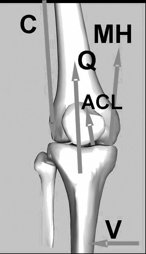
Under external valgus loading, contact shifts to the lateral compartment. The moment balance with respect to the contact point shows that Q and MH both help the ACL (and the medial collateral ligament, not shown) stabilize the joint against valgus loading. Under a given valgus load, any reduction in these muscle forces increases ligament loading
Another imbalance frequently observed in female athletes is quadriceps dominance. Quadriceps dominance is an imbalance between the quadriceps and hamstring recruitment patterns. With quadriceps dominance, female athletes tend to preferentially increase their knee extensor moments over their knee flexor moments when performing sport movements that generate high lower extremity joint torques.22 This overreliance on the quadriceps muscles is hypothesized to lead to imbalances in strength and coordination between the quadriceps and the knee flexor musculature. Hewett et al22 reported that peak flexor moments at the knee were 3-fold higher in male high school athletes than in female athletes landing from a volleyball block. They also demonstrated that peak hamstring torques measured by dynamometry were significantly lower in female athletes than in male athletes. Quadriceps dominance has been observed in elite female collegiate athletes.24 Female athletes reacted to a forward translation of the tibia primarily with a muscular activation of the quadriceps muscles, whereas male athletes relied on their hamstring muscles to counteract the anterior tibial displacement. Malinzak et al39 examined men and women during the sport-specific tasks of running, cross-cutting, and side cutting. Women used less knee flexion, increased quadriceps activation, and decreased hamstring activation compared with men when performing the tasks. This tendency to land with a straighter knee during high-intensity tasks could be exacerbated with premature activation of quadriceps (or delayed activation of hamstring) during weight-bearing stance.40 Chappell et al37 concluded that the increased anterior shear force demonstrated by the female athletes was potentially due to the combination of increased quadriceps force, decreased hamstring force, and decreased knee flexion.
Landing with the knee near full extension is a common mechanism of ACL injury.41 At low knee-flexion angles (0 to 30° of knee flexion), quadriceps contractions pull the tibia forward and increase stress on the ACL, especially without balanced knee-flexor (hamstring and gastrocnemius) cocontraction to decrease strain on the ligament. At knee-flexion angles less than 45°, the quadriceps is an antagonist of the ACL. At knee-flexion angles beyond 45°, the quadriceps translates the tibia in the opposite (posterior) direction, which reduces strain on the ligament.27 Athletes who demonstrate quadriceps dominance may increase their risk for ACL injury when they cut and land with low knee-flexion angles.
Another neuromuscular imbalance that female athletes demonstrate is leg dominance. Leg dominance is the imbalance between muscular strength and joint kinematics in contralateral lower extremity measures. Female athletes have been reported to generate lower hamstring torques in the nondominant than in the dominant leg.22 Ford et al42 showed that adolescent female athletes had significant side-to-side differences in maximum knee-valgus angle compared with male athletes during a box-drop vertical jump. Side-to-side imbalances in muscular strength, flexibility, and coordination have been shown to be important predictors of increased injury risk.23,43,44 Knapik et al44 demonstrated that side-to-side balance in strength and flexibility is important for the prevention of injuries, and when imbalances are present, athletes are more injury prone. Baumhauer et al43 also found that individuals with muscle-strength imbalances exhibited a higher incidence of injury. Hewett et al23 developed a model to predict ACL injury risk with high sensitivity and specificity. Half of the factors in the predictive model were side-to-side differences in lower extremity kinematics and kinetics. Side-to-side imbalances may increase the risk for both limbs. Overreliance on the dominant limb can place greater stress and torques on that knee, whereas the weaker limb is at risk because the musculature cannot effectively absorb the high forces associated with sporting activities.
Neuromuscular imbalances may not be the only factors underlying the sex differences in knee injury rates, but neuromuscular control may be the greatest contributor to dynamic knee stability and offer the greatest potential for intervention.29 Female athletes may especially benefit from neuromuscular training, because they often display decreased baseline levels of strength and power compared with their male counterparts. Comprehensive neuromuscular training programs designed for young women can significantly increase power, strength, and neuromuscular control and decrease sex differences in these measures.45,46 Dynamic neuromuscular training also reduces sex-related differences in force absorption, active joint stabilization, muscle imbalances, and functional biomechanics while increasing the strength of structural tissues (bones, ligaments, and tendons).22,47–50 Most importantly, increasing evidence suggests that different types of neuromuscular training can prevent injuries, specifically ACL injuries.51–53
TRAINING GROUNDWORK
Injury-prevention techniques for the ACL have previously been demonstrated.51–53 Selective incorporation and integration of these techniques could be used to address specific neuromuscular imbalances in female athletes.21 As Griffin reported,29 Henning identified 3 potentially dangerous maneuvers in sport that should be modified through training to prevent ACL injury. He suggested that athletes land in a more bent-knee position and decelerate before a cutting maneuver. Preliminary work implementing these techniques in a pilot sample of athletes suggested a decrease in injury rate in trained versus untrained subjects.29 Subsequently, Hewett et al52 developed a training program, based on a thorough review of the literature and prior athletic training experience, that included an initial phase devoted to correcting jump and landing techniques in female athletes. Four basic techniques were stressed: (1) correct posture (ie, chest over knees) throughout the jump, (2) jumping straight up with no excessive side-to-side or forward-backward movement, (3) soft landings, including toe-to-heel rocking and bent knees, and (4) instant recoil preparation for the next jump. Using the training program, women in the intervention group were able to significantly reduce noncontact ACL injuries compared with controls. Both studies highlight the importance of incorporating dynamic, biomechanically correct movements into training protocols aimed at injury prevention.
Caraffa et al51 prospectively evaluated the effect of balance-board exercises on noncontact ACL injury rates in elite male soccer players. The training consisted of 20 minutes of balance-board exercises divided into 5 phases. Athletes who participated in proprioceptive training before their competitive season had a significantly decreased rate of knee injuries. Conversely, others have examined the effects of similar progressive balance-board training and found no similar reduction in ACL injuries in female athletes.54 Subsequently, Myklebust et al53 examined the effects of a more comprehensive and dynamic neuromuscular training program on female athletes. Their program elaborated on the balance-board protocol of Caraffa et al51 and the techniques of Hewett et al22 by adding a focus to improve awareness and knee control during standing, cutting, jumping, and landing. They were able to reduce the incidence of ACL injury in women's elite handball players over 2 competitive seasons. These studies demonstrate the ability of a neuromuscular/balance component to reduce knee-injury risk in female athletes when incorporated into an injury-prevention protocol.
Education and public awareness of the high rate of occurrence and mechanisms of ACL injury have also been shown to decrease injuries in a group of Vermont ski instructors.55 Ski instructors viewed videotapes of ACL injuries and were encouraged to formulate their own preventive strategies. Pilot data demonstrated a significant reduction in ACL injuries by greater than 50% with this technique. Elements from the Vermont study should be applied to other sports. It is important to teach athletes to avoid biomechanically disadvantageous and dangerous positions in any sport. Hewett et al22 expanded the concept, using clinician verbal and visualization cues to provide feedback and awareness to an athlete during training. Each athlete was encouraged to perform as many jumps as possible using the proper technique. As the athletes became fatigued, they were encouraged to stop if they could not execute each jump with correct biomechanics. Similarly Myklebust et al53 used partner training to provide the critical feedback. Partners encouraged each other to focus on the quality of their movements, specifically on the knee-over-the-toe position. Both Hewett et al22 and Myklebust et al53 recognized the critical analysis as a contributor to the successful reduction of ACL injuries in their studies. Laboratory experiments also provide support for the efficacy of analysis and feedback in reducing dangerous biomechanics. Onate et al56 and Prapavessis and McNair57 reduced peak vertical ground reaction forces using verbal and visual feedback. These groups suggested the inclusion of critical analysis and feedback into injury-prevention models for athletes who participate in sports that require jump landings. Cumulatively, prior studies demonstrated the importance of analyzing the biomechanics of sports movements and providing constant and consistent feedback to the athlete regarding proper technique.
Scientific evidence combined with experience-based empirical evidence allowed us to develop a neuromuscular training rationale. Dynamic neuromuscular analysis training is a synthesis of the most important findings derived from existing research studies and prevention techniques developed through more recent empirical and analytical evaluations of neuromuscular training and on-field play. In contrast to the work of previous authors, dynamic neuromuscular analysis training is not a protocol. Rather, it is a rationalized approach to addressing specific risk factors that may be evident in at-risk athletes. The training principles and techniques provide a general framework to clinicians who wish to design and administer injury-prevention programs targeted to this population.
The 3 essential components of a comprehensive training protocol are dynamic, biomechanically correct movement skills; neuromuscular patterning based on the identification of underlying neuromuscular imbalances; and constant biomechanical analysis by the instructor with feedback to athletes both during and after training. When training to prevent injury, athletes and clinicians must interact. This interactive form of movement training requires intense instructor-to-athlete technique analysis and immediate and consistent feedback, with the goal of programming the neuromuscular system to perform athletic maneuvers in a powerful, efficient, and safe manner.
TRAINING RATIONALE
Dynamic neuromuscular analysis training should address the neuromuscular imbalances present in the population to be trained. Special attention should be given to the female athlete who may display one or more neuromuscular imbalances. Dynamic, multiplanar, sport-specific movements that are a challenge to the proprioceptive system are a required component of neuromuscular analysis training. The exercises selected must challenge the dynamic joint restraints (muscle-tendon units) that maintain limb and joint position in response to changing loads. Dynamic sport-specific training should provide female athletes with an effective means for facilitating the desired adaptations to the proprioceptive function of the knee joint. The dynamic component progresses them to high-risk, sport-specific maneuvers that can be performed in a safe and controlled manner. Properly trained athletes are better prepared both to handle the high joint forces generated during athletic competition in order to reduce the risk of injury and to achieve peak performance.
The neuromuscular component of this training is a balance between challenging an athlete's proprioceptive abilities and exposing her to movement patterns that generate greater dynamic knee control. This proprioceptive stress may aid the development of protective spinal reflexes and multijoint neuromuscular engrams that more effectively manage the ground reaction forces encountered during the midstance phase of high-risk maneuvers. However, assuming that the spinal stretch reflex occurs in 50 to 70 milliseconds, with an additional time lag due to the electromechanical delay, an injury may occur before the reflex response begins to generate significant muscle force at initial contact.58 Neuromuscular training that teaches athletes to better develop joint-stabilization patterns that employ feed-forward mechanisms (muscular preactivation patterns) may preset muscular contraction to increase knee stability at initial contact.59 This enhanced neuromuscular control may help decrease joint motion and protect an athlete's ACL from high impulse loading.58,60
The analysis component of dynamic neuromuscular-analysis training involves exercises that provide instructors with the tools to analyze imperfections in technique. The training should focus on perfecting the technique of each training exercise, especially early in the training cycle. If athletes are allowed to perform the exercise maneuvers improperly, then the training will reinforce improper techniques. Clinicians should give continuous and immediate feedback both during and after each exercise bout to make athletes aware of proper form and technique, as well as undesirable and potentially dangerous positions.57 Additionally, athletes should receive visual feedback via a video camera and television monitor or via exercise in front of a mirror to help make them cognizant of landings with visually identifiable poor biomechanics.56 Visual and verbal feedback helps athletes to match their perceived techniques to their actual techniques. Selected protocol times are general guidelines that provide an expected and attainable goal. Not every athlete will be at a level of muscular strength, coordination, skill, or desire to achieve the selected exercise duration. Clinicians should be skilled in recognizing the desired technique for a given exercise and should learn to encourage athletes to maintain perfect technique for as long as possible. If an athlete fatigues such that she can no longer perform the exercise perfectly and displays a sharp decline in proficiency, then she should be instructed to stop. The duration of each completed exercise should be noted, and the goal of the next training session must be to continue to improve technique and to increase volume (number of repetitions) or intensity (difficulty). The goal of increasing the quantity or intensity of exercises while maintaining the quality of exercises is critical in achieving successful outcomes from the training.
Although prior research demonstrates the benefits of injury-prevention training among a wide variety of athletes, those who demonstrate neuromuscular imbalances as evidenced by poor dynamic knee stability might benefit the most from training.23 The following discussion will address the theoretic basis for the design of a dynamic neuromuscular-analysis training program aimed at correcting specific neuromuscular imbalances in female athletes. The goal of the discussion is to provide clinicians and coaches with specific methods to assess neuromuscular imbalances and to subsequently provide training techniques targeted to correct an athlete's specific imbalances.
IDENTIFICATION OF LIGAMENT DOMINANCE
The measurement of neuromuscular imbalances allows individuals at potential risk for ACL injury to be identified.23 An athlete may display techniques that place her at risk because of one or more imbalances. During single-leg landing, pivoting, or deceleration, the motion of a ligament-dominant knee in an athlete may be directed by the external ground reaction forces, rather than by her musculature.22 Initial evaluation of an athlete's level of ligament dominance can be accomplished using a 31-cm box-drop test combined with a maximum-effort vertical leap.33,49 When landing from a box, a ligament-dominant athlete may display substantial medial knee motion in the coronal plane that can be visually identified. Medial knee motion may be related to an overall dynamic knee valgus (femoral adduction, femoral internal rotation in relation to the hip, tibial external rotation in relation to the femur with or without foot pronation). Movement patterns that place an athlete in positions of high ACL load (excessive external knee-abduction moments) combined with a low knee-flexion angle may increase the risk for ligament injury or failure.61 Landing in the valgus position also correlates with increased impact ground reaction force.22 Thus, a female athlete who lands with excessive medial knee motion may be classified as ligament dominant.
To address ligament dominance, an athlete should be taught to control dynamic knee motion, especially unwanted motions in the coronal plane. She should be shown how to use the knee as a single-plane hinge joint allowing flexion and extension, not valgus and varus. Education for dynamic control of knee motion in the sagittal plane may be achieved through progressive exercises that challenge the neuromuscular system. The first step to addressing ligament dominance is to make an athlete aware of proper form and technique as well as undesirable and potentially dangerous positions. Athletes can be videotaped or placed in front of a mirror to make them aware that they are landing with visually identifiable medial knee motion. Second, clinicians must critically evaluate the jumping and landing sequence and provide constant, technique-oriented feedback. This feedback is similar to the coaching required to teach a specific skill required for a sport.57 Clinicians can use verbal feedback such as “on your toes,” “soft-silent landings,” and “straight as an arrow” as athletes jump; “light as a feather” as they land; and “knees square,” “knees bent,” “shoulders back,” “head up,” and “touch and go.” These cues should be repeated often to help promote a strong foundation of proper technique.22,62 Sports medicine clinicians should attempt to continuously bridge the gap in an athlete's perceived technique and actual technical performance. Clinicians must be diligent in providing adequate feedback for correct technical performance to facilitate the desirable neuromuscular alterations. If the feedback is inadequate or inappropriate, then an athlete may be reinforcing improper techniques with the neuromuscular training.
TREATMENT OF LIGAMENT DOMINANCE
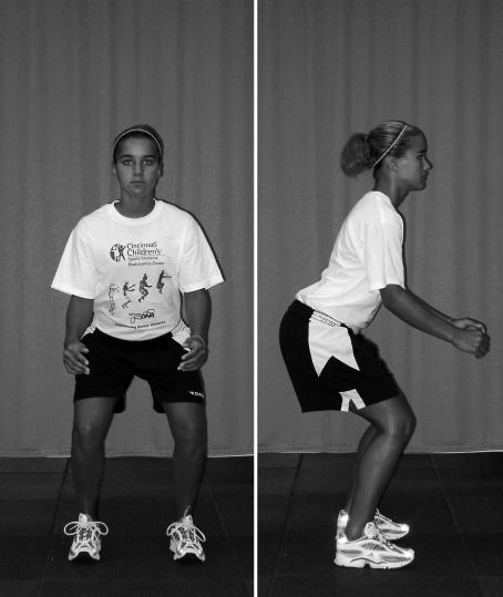
Before being taught the dynamic exercises, athletes should be shown proper athletic position (Figure 2). The athletic position is a functionally stable position with the knees comfortably flexed, shoulders back, eyes up, feet approximately shoulderwidth apart, and body mass balanced over the balls of the feet. The knees should be over the balls of the feet and the chest over the knees.22 This is the athlete-ready position and should be the starting and finishing position for most training exercises.
Figure 2. The athletic position is a functionally stable position with the knees comfortably flexed, shoulders back, eyes up, feet approximately shoulderwidth apart, and body mass balanced over the balls of the feet.
The knees should be over the balls of the feet and the chest over the knees. This athlete-ready position is the starting and finishing position for most of the training exercises. During some exercises, the finishing position is exaggerated with deeper knee flexion in order to emphasize the correction of certain biomechanical deficiencies
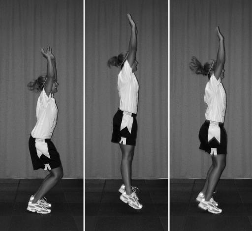
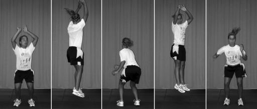
The wall-jump exercise can be used initially to treat ligament dominance (Figure 3). This low- to moderate-intensity jump allows clinicians to begin analyzing an athlete's level of ligament dominance or valgus-varus motion in the knee. During wall jumps, athletes do not go through deep knee-flexion angles; most of the vertical movement is provided by active ankle plantar flexion. The relatively straight knee makes even slight amounts of medial knee motion easy to identify visually. When medial knee motion is observed, clinicians should begin to give verbal feedback cues (eg, “keep knees square,” “no inward knee motion,” “keep knees aligned straight,” “keep knees apart and use your knee like a hinge, not a ball and socket”). This feedback allows athletes to cognitively process the proper knee motion required to perform the exercise; athletes also find this feedback easier to process because the exercise does not require maximal effort. Finally, control of medial knee motion is critical when landing with knee angles close to full extension. Direct ACL load is high during forceful quadriceps contraction with dynamic valgus positioning at small knee-flexion angles.63 Thus, the initial technical goal for a ligament-dominant athlete is to keep the knees apart when landing and to err on the side of varus knee positioning, which produces decreased ACL load in the knee-flexion angles used in this jump.63
Figure 3. Wall jumps.
The athlete stands erect with the arms semiextended overhead. This vertical jump requires minimal knee flexion. The gastrocnemius muscles should create the vertical height, and the arms should extend fully at the top of the jump. Use this jump as a warm-up and coaching exercise, as this relatively low-intensity movement can reveal abnormal knee motion in athletes with poor side-to-side knee control
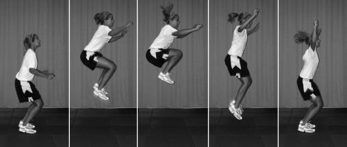
Another useful exercise to target ligament-dominant athletes is the tuck jump (Figure 4). The tuck jump is on the opposite end of the intensity spectrum from the wall jump and requires a high level of effort. Initially, athletes focus most of their cognitive efforts solely on the performance of this difficult jump. Clinicians can quickly identify those athletes who may demonstrate abnormal levels of coronal-plane knee displacement during jumping and landing, because such an athlete usually devotes minimal attention to technique in the first few repetitions. In addition, tuck jumps can be used to assess improvements in lower extremity biomechanics. If an athlete can improve her neuromuscular control and biomechanics during this difficult jumping and landing sequence, then she is gaining dynamic neuromuscular control of the knee joint and may begin to develop a learned skill that might be transferred to competitive play.
Figure 4. Tuck jumps.
The athlete starts in the athletic position with her feet shoulderwidth apart. She initiates the jump with a slight crouch downward while she extends her arms behind her. She then swings her arms forward as she simultaneously jumps straight up and pulls her knees up as high as possible. At the highest point of the jump, the athlete is in the air with her thighs parallel to the ground. When landing, the athlete should immediately begin the next tuck jump. Encourage her to land softly, using a toe-to-midfoot rocker landing. The athlete should not continue this jump if she cannot control the high landing force or if she uses a knock-kneed landing
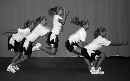
The wall jump and the tuck jump provide clinicians with an analysis of an athlete's movements primarily in the coronal plane. The broad jump and hold (Figure 5) allows clinicians to assess an athlete's knee motion while progressing forward in the sagittal plane. Gaining dynamic knee control while performing multiplanar tasks is critical to changing biomechanics that can transfer to on-the-field play. Some athletes may display active valgus, a position of hip adduction and knee abduction, which is a result of muscular contraction rather than ground reaction forces. Active valgus occurs when taking off from a jump rather than landing and should be corrected during training. The broad jump is a moderate-intensity exercise that offers clinicians another opportunity to provide feedback on technique and to assist athletes to perfect their technique during each jump. In addition, proper execution of the broad jump requires athletes to hold the landing for 3 to 5 seconds, which forces them to gain and maintain dynamic knee control over a more prolonged period of time. By stabilizing her knees over a prolonged period of time, an athlete learns to recognize proper foot positioning and knee control, thereby enhancing proprioceptive and kinesthetic abilities. Recognition of proper positioning and technique is a primary goal of training; however, more reflexive motor patterns are desired for the ultimate success of neuromuscular reprogramming.
Figure 5. Broad jump and hold.
The athlete prepares in the athletic position with her arms extended behind her at the shoulder. She begins by swinging her arms forward and jumping horizontally and vertically at approximately a 45° angle to achieve maximum horizontal distance. The athlete must “stick” the landing with her knees flexed to approximately 90° in an exaggerated athletic position. If she cannot stick the landing during a maximal effort jump in the early phases, have her perform a submaximal broad jump, sticking the landing with her toes straight ahead and no inward motion of her knees. As her technique improves, encourage her to add distance to her jumps but not at the expense of perfect technique
The 180° jump (Figure 6) can be incorporated into training to teach dynamic body and lower extremity control in the transverse plane. The 180° jump creates rotational force in the transverse plane, which must be absorbed and immediately redirected in the opposite direction. This movement is important for teaching an athlete to recognize and control dangerous rotational forces, which if combined with knee abduction or anterior load, produce additive ACL loads.63 Additionally, increased body awareness and control can improve movement patterns, which will reduce injury risk and also improve measures of performance.22,62
Figure 6. The 180° jump.
. The starting position is standing erect with feet shoulderwidth apart. The athlete initiates this 2-footed jump with a direct vertical motion combined with a 180° rotation in midair, keeping her arms away from her sides to help maintain balance. When she lands, she immediately reverses this jump into the opposite direction. She repeats until perfect technique fails. The goal of this jump is to achieve maximal height with a full 180° rotation. Encourage the athlete to maintain exact foot position on the floor by jumping and landing in the same footprint
Once an athlete has learned to jump, land, and hold the broad jump in bipedal stance, the single-leg hop-and-hold exercise (Figure 7) can be incorporated into the training. Proper progression into the single-leg hop and hold is critical to ensure athlete safety during training. This point is salient for clinicians, because ACL injury-prevention techniques should not introduce the inappropriate risk for injury during training. Most noncontact ACL injuries occur when landing or decelerating on a single limb.41 The single-leg hop-and-hold exercise roughly mimics a mechanism of ACL injury during competitive play. When initiating the single-leg hop-and-hold, an athlete should be instructed to jump only a few inches and to land with deep knee flexion. As she masters the low-intensity jumps, the distance can be increased, as long as she can continue to maintain deep knee flexion when landing and control the coronal-plane motion at the knee. Athletes must be given continuous feedback (with cues to “use deep knee flexion,” “sit down deep,” “soft, silent landings,” “light as a feather,” “knee square,” “knee bent”) on both her medial-lateral knee motion control and her ability to absorb the landing in an appropriate manner through ankle, knee, and hip flexion.
Figure 7. Single-leg hop and hold.
The starting position is a semicrouched position on a single leg. The athlete's arm should be fully extended behind her at the shoulder. She initiates the jump by swinging the arms forward while simultaneously extending at the hip and knee. The jump should carry the athlete up at an angle of approximately 45° and attain maximal distance for a single-leg landing. She is instructed to land on the jumping leg with deep knee flexion (to 90°) and to hold the landing for at least 3 seconds. Coach this jump with care to protect the athlete from injury. Start her with a submaximal effort on the single-leg broad jump so she can experience the level of difficulty. Continue to increase the distance of the broad hop as the athlete improves her ability to “stick” and hold the final landing. Have the athlete keep her visual focus away from her feet to help prevent too much forward lean at the waist
The final level of training used to target ligament dominance is unanticipated cutting movements. Before being taught unanticipated cutting, athletes should first be able to proficiently attain the proper athletic position (see Figure 2). This athlete-ready position is the goal position to achieve before initiating a directional cut. Adding the directional cues to the unanticipated part of training can be as simple as pointing or as sport specific as using partner-mimic or ball-retrieval drills.
Single-faceted sagittal-plane training and conditioning protocols that do not incorporate cutting maneuvers will not provide levels of external varus-valgus or rotational loads similar to those seen during sport-specific cutting maneuvers.64 Training programs that incorporate safe levels of varus-valgus stress may induce more muscle-dominant neuromuscular adaptations.61 Such adaptations can better prepare an athlete for more multidirectional sport activities, which can improve performance and reduce the risk of lower extremity injury.22,52,65 Female athletes perform cutting techniques with decreased knee flexion and increased valgus angles.39 Knee-valgus loads can double when an athlete performs unanticipated cutting maneuvers similar to those used in sport.66 Thus, the endpoint of training designed to reduce ACL loading via valgus torques can be achieved by teaching athletes to use movement techniques that produce the low abduction knee-joint moments.61 Recent evidence demonstrates that training incorporating unanticipated movements can reduce knee-joint loads.62 Additionally, training individuals to preactivate their musculature before ground contact may facilitate appropriate kinematic adjustments, and ACL loads may be reduced.66,67 Training an athlete to employ safe cutting techniques in unanticipated sport situations may instill technique adaptations that will more readily transfer onto the field of play. A ligament-dominant athlete may become muscle dominant, reducing her future risk of ACL injury.22,52
IDENTIFICATION OF QUADRICEPS DOMINANCE
Athletes can be screened for indicators of quadriceps dominance using relatively common measurement techniques such as isokinetic dynamometry or perhaps even simple leg-curl and leg-extension machines. Huston and Wojtys24 demonstrated that female athletes had increased quadriceps dominance compared with males and nonathletic controls. If an athlete exhibits a high level of quadriceps strength, a low level of hamstring strength, or a low hamstring-to-quadriceps ratio in one or both limbs, quadriceps dominance may be present. Hamstring-to-quadriceps strength ratios of less than 55% may indicate a quadriceps-dominant athlete who may demonstrate inappropriate or decreased hamstring recruitment patterns during dynamic tasks.68 If objective measures are not available, then an athlete can be tested on her ability to hop and hold a single-leg stance in deep knee flexion. To maintain upright posture with deep knee flexion (>90°) requires relatively more recruitment of hamstrings than quadriceps.69 Inability to maintain stance with deep knee flexion may indicate some level of quadriceps dominance.
TREATMENT OF QUADRICEPS DOMINANCE
To decrease the tendency toward quadriceps dominance, exercises are employed to emphasize cocontraction of the knee flexor-extensor muscles.70 It is difficult to develop a more appropriate firing pattern for the knee flexors while performing exercises that also strongly activate the knee extensors. If the hamstrings are adequately activated at the proper time, they can decrease ACL loading. However, at low knee-flexion angles, the hamstrings have little ability to protect against ACL loads.64,71,72 Additionally, at angles greater than 45°, the quadriceps resist anterior tibial translation, providing an agonistic role to the ACL.73,74 Therefore, it is important to use deep knee-flexion angles to put the quadriceps into an ACL-agonist position and the hamstrings into an ACL-protective position. Athletes trained with deep knee-flexion jumps can learn to increase the amount of knee flexion at landing and decrease the amount of time spent in the more dangerous straight-legged position. We hypothesize that the repetitive achievement of proper positioning may facilitate increased muscle coactivation and possibly lead to reduced ACL loads. When training female athletes with dynamic exercises, especially exercises that use deep knee-flexion angles, clinicians should be aware of the potential introduction of anterior knee pain, which may occur at an increased rate in this population.75 Modifying the jump exercises with decreased knee flexion and pain-free range of motion may be warranted for training athletes to reduce the potential for patellofemoral pain.76
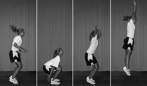
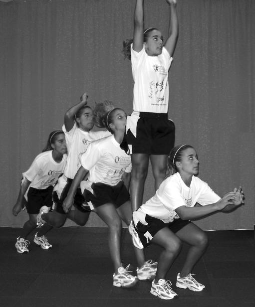
Squat jumps (Figure 8) and broad jump and holds (see Figure 5) can specifically address the training goal of improving the protective nature of increased sagittal-plane flexor moments. Squat jumps require an athlete to go into deep knee-flexion angles, past 90°. Squat jumps may increase the relative recruitment and strength of the flexor musculature, as well as provide a mechanism to learn closed-chain knee control over a large range of motion.69 Additionally, the squat jump can help teach an athlete to land in a more flexed-knee position, which decreases the quadriceps' ability to load the ACL and improves the ability of the hamstrings to offset anterior shear forces due to their line of pull.64,71–74
Figure 8. Squat jumps.
The athlete begins in the athletic position with her feet flat on the mat and pointing straight ahead. She drops into deep knee, hip, and ankle flexion; touches the floor (or mat) as close to her heels as possible; and then takes off into a maximal vertical jump. The athlete then jumps straight up vertically and reaches as high as possible. On landing, she immediately returns to the starting position and repeats the initial jump. Repeat for the allotted time or until her technique begins to deteriorate. Teach the athlete to jump straight up vertically, reaching as high overhead as possible. Encourage her to land in the same spot on the floor and maintain upright posture when regaining the deep-squat position. Do not allow the athlete to bend forward at the waist to reach the floor. She should keep her eyes up, feet and knees pointed straight ahead, and arms to the outside of her legs
Although large knee-flexion angle jumps, like squat jumps, may provide increased hamstring activation through continuous knee range of motion, the broad jump and hop-and-hold exercises (see Figure 5) are important for training the hamstring cocontraction to provide stabilization in static position. When performing the broad-jump-and-hold exercise, hamstring firing is required early in the landing to prevent the anterior tibial shear force needed to counteract the quadriceps firing during deceleration from a landing and later in the maneuver to prevent the dangerous valgus knee motion. Hamstring muscles work to provide resistance against these high force motions, but they must also provide adequate cocontraction to maintain upright posture. Thus, repetitive training with deep knee-flexion hold exercises may improve hamstring strength and recruitment and quadriceps agonistic support and reinforce safe positioning when performing sport maneuvers.
IDENTIFICATION OF LEG DOMINANCE
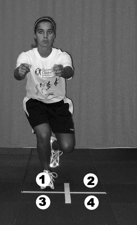
Leg dominance can be assessed using a dynamometer or leg-curl and leg-extension machines. A difference in strength or power of 20% or more between limbs indicates a neuromuscular imbalance that may underlie significant injury risk.44 Another indicator of bilateral imbalances is an athlete's ability to perform a single-leg balanced stance on an unbalanced platform that can objectively quantify postural sway (ie, a stabilometer). Women demonstrate poor scores on stabilometry measures taken on unaffected limbs after ACL injury.31 These pilot data may indicate some relationship between ACL injury risk and poor stabilometric scores. Tropp and Odenrick77 reported that athletes who could not demonstrate postural balance within 2 standard deviations of normal had a significantly higher risk for an ankle injury. Increased proficiency in bipedal and single-leg balance can be gained through balance-board training.78 Tropp and Odenrick77 were able to reduce injury rates in athletes returning to sport from prior injury with 10 weeks of balance-board training. Finally, field exercises such as X hops (Figure 9) can be used as a measure to grade bilateral differences in single-limb performance. Identifying limb imbalances will assist clinicians and researchers in intervening with athletes who need dynamic neuromuscular analysis training.
Figure 9. X hops.
The athlete faces a quadrant pattern and stands on a single limb with the support knee slightly bent. She hops diagonally, lands in the opposite quadrant, maintains forward stance, and holds the deep knee-flexion landing for 3 seconds. She then hops laterally into the side quadrant and again holds the landing. Next she hops diagonally backward and holds the jump. Finally, she hops laterally into the initial quadrant and holds the landing. She repeats this pattern for the required number of sets. Encourage the athlete to maintain balance during each landing, keeping her eyes up and the visual focus away from her feet
TREATMENT OF LEG DOMINANCE
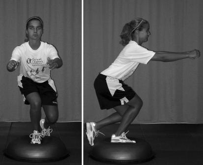
In order to correct for leg dominance, training must progressively emphasize double-leg and then single-leg movements. Equal leg-to-leg strength, balance, and foot placement are stressed throughout the program. For example, to perform tuck jumps (see Figure 3), a leg-dominant athlete may repeatedly place the weaker limb under greater stress to maintain symmetry throughout the performance of the double-leg jump. If she does not produce equal force output in each leg, then she will have difficulty maintaining proper position in jumping and landing and will have increased difficulty maintaining an upright vertical tuck jump. Thus, to perform the tuck jump, the weaker leg must work harder to maintain an equal position with the stronger leg. In turn, greater neuromuscular adaptation may be evoked in the weaker limb. An important training point for clinicians is to pay special attention to foot placement during landing and jumping. Athletes should not be allowed to drop one leg posterior to the other leg when performing double-leg jumps. This position will only facilitate leg-to-leg imbalances further by overloading the stronger (posteriorly positioned) leg, while unloading the stress on the weaker (anteriorly positioned) leg. An athlete who continuously exhibits the problem of dropping a leg or overusing the stronger leg can reprogram this dangerous pattern with single-leg exercises such as the hop-and-hold exercise mentioned earlier or with the single-leg balance exercise on an unstable surface (Figure 10). By forcing each limb to work independently of the other limb, no compensation can be provided by the contralateral limb. Similar amounts of work force greater muscular adaptations in the weaker limb and may decrease imbalances that may have been displayed before training.
Figure 10. Single-leg balance.
The balance drills are performed on a balance device that provides an unstable surface. The athlete begins on the device with a 2-legged stance with feet shoulderwidth apart, in athletic position. As she improves, the training drills can incorporate ball catches and single-leg balance drills. Encourage the athlete to maintain deep knee flexion when performing all balance drills
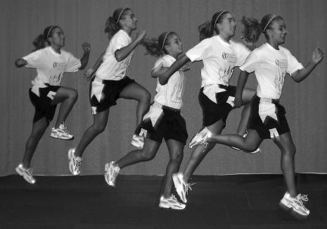
Bounding exercises (Figure 11) can be incorporated into training to aid in the correction of lower extremity imbalances and to help an athlete learn to coordinate multiplanar movements. To correctly perform the bounding exercise, an athlete should jump with maximum distance in both the vertical and horizontal planes. This power jump incorporates high-intensity jumps and landings on a single limb. This type of high-intensity, single-leg movement can accelerate the correction of imbalances between the lower extremities. During this exercise, the weaker leg is repeatedly and quickly exposed to the forces generated by the other leg. The neuromuscular stress placed on the less coordinated leg to individually dampen and quickly redirect the forces generated by the dominant limb during this exercise may help to normalize any bilateral strength or coordination discrepancies between the lower extremities.
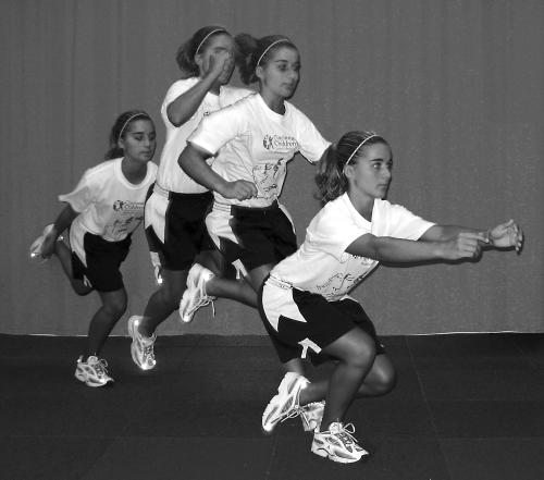
Figure 11. Bounding.
The athlete begins this jump by bounding in place. Once she attains proper rhythm and form, encourage her to maintain the vertical component of the bound while adding some horizontal distance to each jump. The progression of jumps advances the athlete across the training area. When coaching this jump, encourage the athlete to maintain maximum bounding height
Correction of leg dominance requires an athlete to learn to coordinate multijoint actions and multiplanar movements into power skill movements that can be used during competitive play. The endpoint of training should incorporate exercises that force an athlete to use maximum effort with perfect technique. The jump, jump, jump, vertical jump (Figure 12) can be used to evaluate an athlete's potential ability to transfer proper techniques into sport-related tasks. This exercise is performed with maximum effort for horizontal momentum, which must quickly and efficiently be transferred into vertical movement. Execution of this movement is similar to that during competitive play. Examples may include a soccer player who must rapidly stop and jump to head the ball, a basketball player performing a jump-stop layup, or a volleyball player who uses the approach to gain momentum for maximum height during the attack. An improper or unbalanced limb in any step of the jump sequence makes the jump difficult to execute. The reward of a more easily accomplished task pushes an athlete to learn bilateral limb control during this jump.
Figure 12. Jump, jump, jump, vertical jump.
The athlete performs 3 successive broad jumps and immediately progresses into a maximum-effort vertical jump. The 3 consecutive broad jumps should be performed as quickly as possible and attain maximal horizontal distance. The third broad jump should be used as a preparatory jump that will allow horizontal momentum to be quickly and efficiently transferred into vertical power. Encourage the athlete to provide minimal braking on the third and final broad jump to ensure that maximum energy is transferred to the vertical jump. Coach the athlete to go directly vertical on the fourth jump and not move horizontally. Use full arm extension to achieve maximum vertical height
In summary, the progression from double-leg to single-leg power maneuvers is a requirement for correcting leg-to-leg imbalances. In addition, the incorporation of multiplanar movements that equally recruit both lower extremities for optimal performance is necessary. More complex movement patterns require greater synchronization and coordination in side-to-side performance, which leads to greater balance in side-to-side muscle recruitment and equalization of leg-to-leg coordination and power.
CONCLUSIONS
Female athletes may demonstrate one or more neuromuscular imbalances of ligament dominance, quadriceps dominance, and leg dominance. Dynamic neuromuscular analysis training provides a method to specifically address and correct these neuromuscular imbalances. Correction of neuromuscular imbalances is important for both optimal biomechanics of athletic movements and reduction in knee injuries. Objective, standardized jump-landing risk assessments that can be quickly and easily administered by clinicians should be encouraged for preseason screening of potentially at-risk female athletes. Further study on the effects of neuromuscular retraining on biomechanical performance and knee-injury incidence is important to advance injury-prevention programs and women's athletics. Although significant strides have been made, continued advancements in sports injury-risk screening, including the incorporation of high-risk sports movements and maturational assessment into the screening tests, are necessary. Furthermore, research is warranted to determine the most effective timing for interventional training in young female athletes, with the goals of maximizing efficiency and precluding them from high-risk competitive seasons.
Acknowledgments
We acknowledge Ton Van den Bogert of The Cleveland Clinic Foundation for his assistance with Figure 1. We also thank all the athletes and their schools in the greater Cincinnati area who participated in the studies. We recognize Dr. Jeffrey Robbins, Director of The Division of Molecular Cardiovascular Biology, Cincinnati Children's Medical Center, for his invaluable support. We would like to acknowledge funding support from National Institutes of Health Grant R01-AR049735-01A1(TEH).
REFERENCES
- Frank CB, Jackson DW. The science of reconstruction of the anterior cruciate ligament. J Bone Joint Surg Am. 1997;79:1556–1576. doi: 10.2106/00004623-199710000-00014. [DOI] [PubMed] [Google Scholar]
- Fleming BC. Biomechanics of the anterior cruciate ligament. J Orthop Sports Phys Ther. 2003;33:A13–A15. [PubMed] [Google Scholar]
- Arendt E, Dick R. Knee injury patterns among men and women in collegiate basketball and soccer: NCAA data and review of literature. Am J Sports Med. 1995;23:694–701. doi: 10.1177/036354659502300611. [DOI] [PubMed] [Google Scholar]
- Malone TR, Hardaker WT, Garrett WE, Feagin JA, Bassett FH. Relationship of gender to anterior cruciate ligament injuries in intercollegiate basketball players. J South Orthop Assoc. 1993;2:36–39. [Google Scholar]
- National Federation of State High School Associations. Indianapolis, IN: National Federation of State High School Associations; 2002. 2002 High School Participation Survey. [Google Scholar]
- National Collegiate Athletic Association. Indianapolis, IN: National Collegiate Athletic Association; 2002. NCAA Injury Surveillance System Summary. [Google Scholar]
- Toth AP, Cordasco FA. Anterior cruciate ligament injuries in the female athlete. J Gend Specif Med. 2001;4:25–34. [PubMed] [Google Scholar]
- Hewett TE, Lindenfeld TN, Riccobene JV, Noyes FR. The effect of neuromuscular training on the incidence of knee injury in female athletes: a prospective study. Am J Sports Med. 1999;27:699–706. doi: 10.1177/03635465990270060301. [DOI] [PubMed] [Google Scholar]
- Freedman KB, Glasgow MT, Glasgow SG, Bernstein J. Anterior cruciate ligament injury and reconstruction among university students. Clin Orthop. 1998;356:208–212. doi: 10.1097/00003086-199811000-00028. [DOI] [PubMed] [Google Scholar]
- Deacon A, Bennell K, Kiss ZS, Crossley K, Brukner P. Osteoarthritis of the knee in retired, elite Australian Rules footballers. Med J Aust. 1997;166:187–190. doi: 10.5694/j.1326-5377.1997.tb140072.x. [DOI] [PubMed] [Google Scholar]
- Andrish JT. Anterior cruciate ligament injuries in the skeletally immature patient. Am J Orthop. 2001;30:103–110. [PubMed] [Google Scholar]
- Buehler-Yund C. Environmental Health. Cincinnati, OH: University of Cincinnati; 1999. A longitudinal study of injury rates and risk factors in 5 to 12 year old soccer players; p. 161. [Google Scholar]
- Clanton TO, DeLee JC, Sanders B, Neidre A. Knee ligament injuries in children. J Bone Joint Surg Am. 1979;61:1195–1201. [PubMed] [Google Scholar]
- Gallagher SS, Finison K, Guyer B, Goodenough S. The incidence of injuries among 87,000 Massachusetts children and adolescents: results of the 1980–81 Statewide Childhood Injury Prevention Program Surveillance System. Am J Public Health. 1984;74:1340–1347. doi: 10.2105/ajph.74.12.1340. [DOI] [PMC free article] [PubMed] [Google Scholar]
- Tursz A, Crost M. Sports-related injuries in children: a study of their characteristics, frequency, and severity, with comparison to other types of accidental injuries. Am J Sports Med. 1986;14:294–299. doi: 10.1177/036354658601400409. [DOI] [PubMed] [Google Scholar]
- Beunen G, Malina RM. Growth and physical performance relative to the timing of the adolescent spurt. Exerc Sport Sci Rev. 1988;16:503–540. [PubMed] [Google Scholar]
- Malina RM, Bouchard C. Growth, Maturation, and Physical Activity. Champaign, IL: Human Kinetics; 1991. Timing and sequence of changes in growth, maturation, and performance during adolescence; pp. 267–272. [Google Scholar]
- Kellis E, Tsitskaris GK, Nikopoulou MD, Moiusikou KC. The evaluation of jumping ability of male and female basketball players according to their chronological age and major leagues. J Strength Cond Res. 1999;13:40–46. [Google Scholar]
- Malina RM, Bouchard C. Champaign, IL: Human Kinetics; 1991. Growth, Maturation, and Physical Activity; p. xiii,501. [Google Scholar]
- Martin RJ, Dore E, Twisk J, Van Praagh E, Hautier CA, Bedu M. Longitudinal changes of maximal short-term peak power in girls and boys during growth. Med Sci Sports Exerc. 2004;36:498–503. doi: 10.1249/01.mss.0000117162.20314.6b. [DOI] [PubMed] [Google Scholar]
- Hewett TE, Myer GD, Ford KR. Prevention of anterior cruciate ligament injuries. Curr Womens Health Rep. 2001;1:218–224. [PubMed] [Google Scholar]
- Hewett TE, Stroupe AL, Nance TA, Noyes FR. Plyometric training in female athletes: decreased impact forces and increased hamstring torques. Am J Sports Med. 1996;24:765–773. doi: 10.1177/036354659602400611. [DOI] [PubMed] [Google Scholar]
- Hewett TE, Myer GD, Ford KR, et al. Neuromuscular control and valgus loading of the knee predict ACL injury risk in female athletes. Am J Sports Med. doi: 10.1177/0363546504269591. In press. [DOI] [PubMed] [Google Scholar]
- Huston LJ, Wojtys EM. Neuromuscular performance characteristics in elite female athletes. Am J Sports Med. 1996;24:427–436. doi: 10.1177/036354659602400405. [DOI] [PubMed] [Google Scholar]
- Beynnon BD, Fleming BC. Anterior cruciate ligament strain in-vivo: a review of previous work. J Biomech. 1998;31:519–525. doi: 10.1016/s0021-9290(98)00044-x. [DOI] [PubMed] [Google Scholar]
- Li G, Rudy TW, Sakane M, Kanamori A, Ma CB, Woo SL. The importance of quadriceps and hamstring muscle loading on knee kinematics and in-situ forces in the ACL. J Biomech. 1999;32:395–400. doi: 10.1016/s0021-9290(98)00181-x. [DOI] [PubMed] [Google Scholar]
- Markolf KL, Graff-Radford A, Amstutz HC. In vivo knee stability: a quantitative assessment using an instrumented clinical testing apparatus. J Bone Joint Surg Am. 1978;60:664–674. [PubMed] [Google Scholar]
- Besier TF, Lloyd DG, Cochrane JL, Ackland TR. External loading of the knee joint during running and cutting maneuvers. Med Sci Sports Exerc. 2001;33:1168–1175. doi: 10.1097/00005768-200107000-00014. [DOI] [PubMed] [Google Scholar]
- Griffin LY. Prevention of Noncontact ACL Injuries. Rosemont, IL: American Academy of Orthopaedic Surgeons; 2001. The Henning program; pp. 93–96. [Google Scholar]
- Hewett TE. Preventing Knee Injuries in Female Athletes: Tenth Annual Mayo Clinic Symposium on Sports Medicine. Rochester, MN: The Mayo Clinic; 2000. Nov 17–18, Preventing knee injuries in female athletes; pp. 45–55. [Google Scholar]
- Hewett TE, Paterno MV, Myer GD. Strategies for enhancing proprioception and neuromuscular control of the knee. Clin Orthop. 2002;402:76–94. doi: 10.1097/00003086-200209000-00008. [DOI] [PubMed] [Google Scholar]
- Andrews JR, Axe MJ. The classification of knee ligament instability. Orthop Clin North Am. 1985;16:69–82. [PubMed] [Google Scholar]
- Ford KR, Myer GD, Hewett TE. Valgus knee motion during landing in high school female and male basketball players. Med Sci Sports Exerc. 2003;35:1745–1750. doi: 10.1249/01.MSS.0000089346.85744.D9. [DOI] [PubMed] [Google Scholar]
- Myer GD, Ford KR, Hewett TE. A comparison of medial knee motion in basketball players when performing a basketball rebound [abstract] Med Sci Sports Exerc. 2002;34:S5. [Google Scholar]
- Winter DA. New York, NY: John Wiley & Sons; 1990. Biomechanics and Motor Control of Human Movement. 2nd ed; pp. 41–45. [Google Scholar]
- Ireland ML. The female ACL: why is it more prone to injury? Orthop Clin North Am. 2002;33:637–651. doi: 10.1016/s0030-5898(02)00028-7. [DOI] [PubMed] [Google Scholar]
- Chappell JD, Yu B, Kirkendall DT, Garrett WE. A comparison of knee kinetics between male and female recreational athletes in stop-jump tasks. Am J Sports Med. 2002;30:261–267. doi: 10.1177/03635465020300021901. [DOI] [PubMed] [Google Scholar]
- Hewett TE, Myer GD, Ford KR. Decrease in neuromuscular control about the knee with maturation in female athletes. J Bone Joint Surg Am. 2004;86:1601–1608. doi: 10.2106/00004623-200408000-00001. [DOI] [PubMed] [Google Scholar]
- Malinzak RA, Colby SM, Kirkendall DT, Yu B, Garrett WE. A comparison of knee joint motion patterns between men and women in selected athletic tasks. Clin Biomech (Bristol, Avon) 2001;16:438–445. doi: 10.1016/s0268-0033(01)00019-5. [DOI] [PubMed] [Google Scholar]
- Shultz SJ, Perrin DH, Adams MJ, Arnold BL, Gansneder BM, Granata KP. Neuromuscular response characteristics in men and women after knee perturbation in a single-leg, weight-bearing stance. J Athl Train. 2001;36:37–43. [PMC free article] [PubMed] [Google Scholar]
- Boden BP, Dean GS, Feagin JA, Garrett WE. Mechanisms of anterior cruciate ligament injury. Orthopedics. 2000;23:573–578. doi: 10.3928/0147-7447-20000601-15. [DOI] [PubMed] [Google Scholar]
- Ford KR, Myer GD, Hewett TE. Valgus knee motion during landing in high school female and male basketball players. Med Sci Sports Exerc. 2003;35:1745–1750. doi: 10.1249/01.MSS.0000089346.85744.D9. [DOI] [PubMed] [Google Scholar]
- Baumhauer JF, Alosa DM, Renstrom AR, Trevino S, Beynnon B. A prospective study of ankle injury risk factors. Am J Sports Med. 1995;23:564–570. doi: 10.1177/036354659502300508. [DOI] [PubMed] [Google Scholar]
- Knapik JJ, Bauman CL, Jones BH, Harris JM, Vaughan L. Preseason strength and flexibility imbalances associated with athletic injuries in female collegiate athletes. Am J Sports Med. 1991;19:76–81. doi: 10.1177/036354659101900113. [DOI] [PubMed] [Google Scholar]
- Kraemer WJ, Hakkinen K, Triplett-Mcbride NT, et al. Physiological changes with periodized resistance training in women tennis players. Med Sci Sports Exerc. 2003;35:157–168. doi: 10.1097/00005768-200301000-00024. [DOI] [PubMed] [Google Scholar]
- Kraemer WJ, Mazzetti SA, Nindl BC, et al. Effect of resistance training on women's strength/power and occupational performances. Med Sci Sports Exerc. 2001;33:1011–1025. doi: 10.1097/00005768-200106000-00022. [DOI] [PubMed] [Google Scholar]
- Faigenbaum AD, Kraemer WJ, Cahill B, et al. Youth resistance training: position statement paper and literature review. Strength Cond. 1996 Dec;:62–75. [Google Scholar]
- Fleck SJ, Falkel JE. Value of resistance training for the reduction of sports injuries. Sports Med. 1986;3:61–68. doi: 10.2165/00007256-198603010-00006. [DOI] [PubMed] [Google Scholar]
- Myer GD, Hewett TE, Noyes FR. The use of video analysis to identify athletes with increased valgus knee excursion: effects of gender and training [abstract] Med Sci Sports Exerc. 2000;32:S298. [Google Scholar]
- Rooks DS, Micheli LJ. Musculoskeletal assessment and training: the young athlete. Clin Sports Med. 1988;7:641–677. [PubMed] [Google Scholar]
- Caraffa A, Cerulli G, Projetti M, Aisa G, Rizzo A. Prevention of anterior cruciate ligament injuries in soccer: a prospective controlled study of proprioceptive training. Knee Surg Sports Traumatol Arthrosc. 1996;4:19–21. doi: 10.1007/BF01565992. [DOI] [PubMed] [Google Scholar]
- Hewett TE, Riccobene JV, Lindenfeld TN, Noyes FR. The effect of neuromuscular training on the incidence of knee injury in female athletes: a prospective study. Am J Sports Med. 1999;27:699–706. doi: 10.1177/03635465990270060301. [DOI] [PubMed] [Google Scholar]
- Myklebust G, Engebretsen L, Braekken IH, Skjolberg A, Olsen OE, Bahr R. Prevention of anterior cruciate ligament injuries in female team handball players: a prospective intervention study over three seasons. Clin J Sport Med. 2003;13:71–78. doi: 10.1097/00042752-200303000-00002. [DOI] [PubMed] [Google Scholar]
- Soderman K, Werner S, Pietila T, Engstrom B, Alfredson H. Balance board training: prevention of traumatic injuries of the lower extremities in female soccer players? A prospective randomized intervention study. Knee Surg Sports Traumatol Arthrosc. 2000;8:356–363. doi: 10.1007/s001670000147. [DOI] [PubMed] [Google Scholar]
- Johnson RJ. The ACL injury in female skiers. In: Griffin LY, editor. Prevention of Noncontact ACL Injuries. Rosemont, IL: American Academy of Orthopaedic Surgeons; 2001. pp. 107–111. [Google Scholar]
- Onate JA, Guskiewicz KM, Sullivan RJ. Augmented feedback reduces jump landing forces. J Orthop Sports Phys Ther. 2001;31:511–517. doi: 10.2519/jospt.2001.31.9.511. [DOI] [PubMed] [Google Scholar]
- Prapavessis H, McNair PJ. Effects of instruction in jumping technique and experience jumping on ground reaction forces. J Orthop Sports Phys Ther. 1999;29:352–356. doi: 10.2519/jospt.1999.29.6.352. [DOI] [PubMed] [Google Scholar]
- Solomonow M, Krogsgaard M. Sensorimotor control of knee stability: a review. Scand J Med Sci Sports. 2001;11:64–80. doi: 10.1034/j.1600-0838.2001.011002064.x. [DOI] [PubMed] [Google Scholar]
- Rapoport S, Mizrahi J, Kimmel E, Verbitsky O, Isakov E. Constant and variable stiffness and damping of the leg joints in human hopping. J Biomech Eng. 2003;125:507–514. doi: 10.1115/1.1590358. [DOI] [PubMed] [Google Scholar]
- Lloyd DG, Buchanan TS. A model of load sharing between muscles and soft tissues at the human knee during static tasks. J Biomech Eng. 1996;118:367–376. doi: 10.1115/1.2796019. [DOI] [PubMed] [Google Scholar]
- Lloyd DG. Rationale for training programs to reduce anterior cruciate ligament injuries in Australian football. J Orthop Sports Phys Ther. 2001;31:645–654661. doi: 10.2519/jospt.2001.31.11.645. [DOI] [PubMed] [Google Scholar]
- Myer GD, Ford KR, Palumbo JP, Hewett TE. Comprehensive neuromuscular training improves both performance and lower extremity biomechanics in female athletes. J Strength Cond Res. doi: 10.1519/13643.1. In press. [DOI] [PubMed] [Google Scholar]
- Markolf KL, Burchfield DM, Shapiro MM, Shepard MF, Finerman GA, Slauterbeck JL. Combined knee loading states that generate high anterior cruciate ligament forces. J Orthop Res. 1995;13:930–935. doi: 10.1002/jor.1100130618. [DOI] [PubMed] [Google Scholar]
- Lloyd DG, Buchanan TS. Strategies of muscular support of varus and valgus isometric loads at the human knee. J Biomech. 2001;34:1257–1267. doi: 10.1016/s0021-9290(01)00095-1. [DOI] [PubMed] [Google Scholar]
- Cahill BR, Griffith EH. Effect of preseason conditioning on the incidence and severity of high school football knee injuries. Am J Sports Med. 1978;6:180–184. doi: 10.1177/036354657800600406. [DOI] [PubMed] [Google Scholar]
- Besier TF, Lloyd DG, Ackland TR, Cochrane JL. Anticipatory effects on knee joint loading during running and cutting maneuvers. Med Sci Sports Exerc. 2001;33:1176–1181. doi: 10.1097/00005768-200107000-00015. [DOI] [PubMed] [Google Scholar]
- Neptune RR, Wright IC, van den Bogert AJ. Muscle coordination and function during cutting movements. Med Sci Sports Exerc. 1999;31:294–302. doi: 10.1097/00005768-199902000-00014. [DOI] [PubMed] [Google Scholar]
- Davies G. Onalaska, WI: S&S Publishers; 1992. Compendium of Isokinetics in Clinical Usage. 4th ed. [Google Scholar]
- Caterisano A, Moss RF, Pellinger TK, et al. The effect of back squat depth on the EMG activity of 4 superficial hip and thigh muscles. J Strength Cond Res. 2002;16:428–432. [PubMed] [Google Scholar]
- Fitzgerald GK, Axe MJ, Snyder-Mackler L. Proposed practice guidelines for nonoperative anterior cruciate ligament rehabilitation of physically active individuals. J Orthop Sports Phys Ther. 2000;30:194–203. doi: 10.2519/jospt.2000.30.4.194. [DOI] [PubMed] [Google Scholar]
- More RC, Karras BT, Neiman F, Fritschy D, Woo SL-Y, Daniel DM. Hamstrings, an anterior cruciate ligament protagonist: an in vitro study. Am J Sports Med. 1993;21:231–237. doi: 10.1177/036354659302100212. [DOI] [PubMed] [Google Scholar]
- Pandy MG, Shelburne KB. Dependence of cruciate-ligament loading on muscle forces and external load. J Biomech. 1997;30:1015–1024. doi: 10.1016/s0021-9290(97)00070-5. [DOI] [PubMed] [Google Scholar]
- Andriacchi TP, Andersson GBJ, Fermier RW, Stern D, Galante JO. A study of lower-limb mechanics during stair-climbing. J Bone Joint Surg Am. 1980;62:749–757. [PubMed] [Google Scholar]
- Daniel DM, Malcom LL, Losse G, Stone ML, Sachs R, Burks R. Instrumented measurement of anterior laxity of the knee. J Bone Joint Surg Am. 1985;67:720–726. [PubMed] [Google Scholar]
- DeHaven KE, Lintner DM. Athletic injuries: comparison by age, sport, and gender. Am J Sports Med. 1986;14:218–224. doi: 10.1177/036354658601400307. [DOI] [PubMed] [Google Scholar]
- Richards DP, Ajemian SV, Wiley JP, Zernicke RF. Knee joint dynamics predict patellar tendinitis in elite volleyball players. Am J Sports Med. 1996;24:676–683. doi: 10.1177/036354659602400520. [DOI] [PubMed] [Google Scholar]
- Tropp H, Odenrick P. Postural control in single-limb stance. J Orthop Res. 1988;6:833–839. doi: 10.1002/jor.1100060607. [DOI] [PubMed] [Google Scholar]
- Tropp H, Ekstrand J, Gillquist J. Stabilometry in functional instability of the ankle and its value in predicting injury. Med Sci Sports Exerc. 1984;16:64–66. [PubMed] [Google Scholar]


