An 83-year-old woman presented to our emergency department with a two-week history of progressive dyspnoea on exertion and leg oedema, and no syncope. She had a history of hypertension, diabetes mellitus type 2, renal insufficiency, and left bundle branch block.
She was clinically mildly decompensated. The ECG showed sinus rhythm with total AV block and a ventricular escape rhythm of 30/min. Echocardiography showed normal left and right ventricular systolic function and a dilated inferior vena cava with decreased variation.
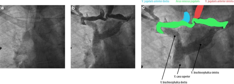
During pacemaker implantation, a remarkable anatomy of the jugular veins was noticed. An aberrant trajectory of the wire was perceived from the left subclavian vein to the contralateral side (Fig. 1a and online video). After contrast injection, two large veins were observed running parallel to one another which were connected caudally (Fig. 1b). Furthermore, the left brachiocephalic vein and superior vena cava (Fig. 1b and 1c) are appreciated.
Fig. 1.
a Chest radiography showing a guide wire (arrow head) running horizontally from the left subclavian vein to the contralateral side. b An overlay of sequential chest X‑rays showing a phlebogram of two anterior jugular veins running parallel (venae jugulares anteriores) which are connected caudally by a jugular arch (arcus venosus juguli). See also the online video
We concluded that the anatomy consisted of enlarged anterior jugular veins (venae jugulares anteriores) and a jugular arch (arcus venosus juguli) [1]. This is a common anatomy, but these jugular veins are rarely enlarged [2]. Probably, this has to do with the increased venous pressure due to backward heart failure.
Caption Electronic Supplementary Material
Video of serial contrast injections showing flebograms of two anterior jugular veins (venae jugulares anteriores) running parallel which are connected caudally by a jugular arch (arcus venosus juguli). See also Fig. 1.
Acknowledgments
Acknowledgements
We thank Dr. Marc Vorstenbosch, Associate Professor at the Department of Anatomy of the Radboud University Medical Center, Nijmegen, the Netherlands, for his critical appraisal of this manuscript.
Conflict of interest
M. Boulaksil and R.M.M. Gevers declare that they have no competing interests.
References
- 1.Schummer W, Schummer C, Bredle D, Frober R. The anterior jugular venous system: variability and clinical impact. Anesth Analg. 2004;99:1625–1629. doi: 10.1213/01.ANE.0000138038.33738.32. [DOI] [PubMed] [Google Scholar]
- 2.Nayak BS. Surgically important variations of the jugular veins. Clin Anat. 2006;19:544–546. doi: 10.1002/ca.20268. [DOI] [PubMed] [Google Scholar]
Associated Data
This section collects any data citations, data availability statements, or supplementary materials included in this article.
Supplementary Materials
Video of serial contrast injections showing flebograms of two anterior jugular veins (venae jugulares anteriores) running parallel which are connected caudally by a jugular arch (arcus venosus juguli). See also Fig. 1.


