

Abstract
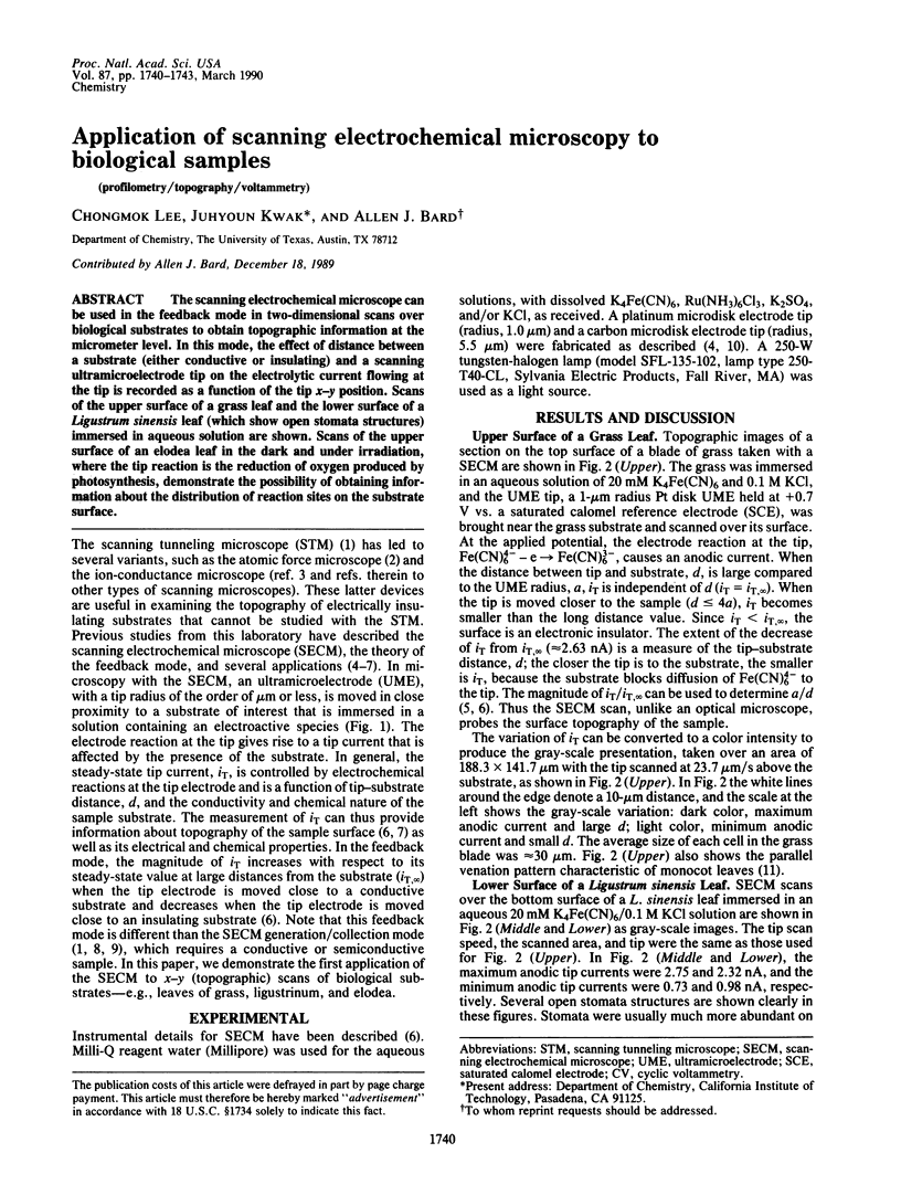
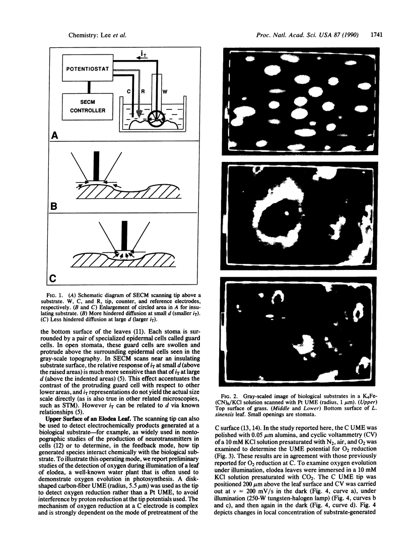
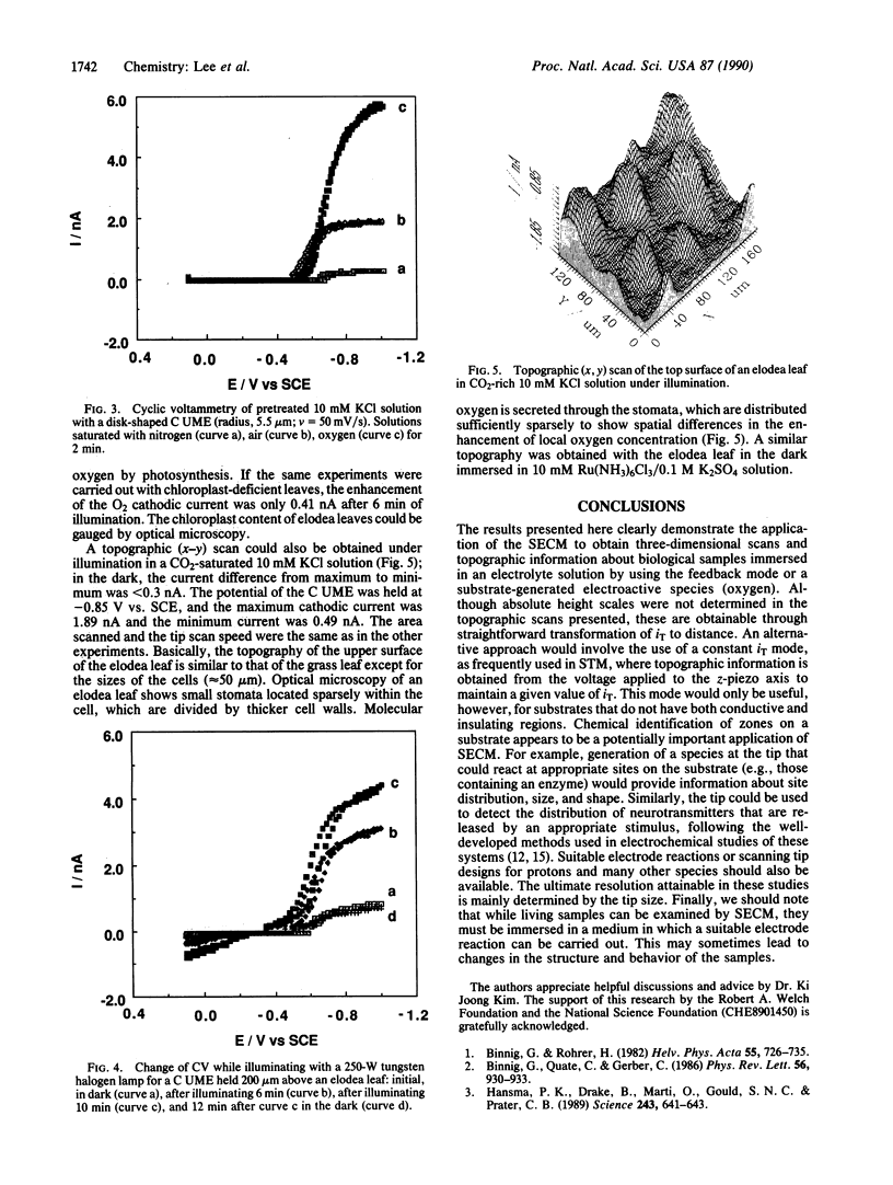
The scanning electrochemical microscope can be used in the feedback mode in two-dimensional scans over biological substrates to obtain topographic information at the micrometer level. In this mode, the effect of distance between a substrate (either conductive or insulating) and a scanning ultramicroelectrode tip on the electrolytic current flowing at the tip is recorded as a function of the tip x-y position. Scans of the upper surface of a grass leaf and the lower surface of a Ligustrum sinensis leaf (which show open stomata structures) immersed in aqueous solution are shown. Scans of the upper surface of an elodea leaf in the dark and under irradiation, where the tip reaction is the reduction of oxygen produced by photosynthesis, demonstrate the possibility of obtaining information about the distribution of reaction sites on the substrate surface.
Full text
PDF

Images in this article
Selected References
These references are in PubMed. This may not be the complete list of references from this article.
- Adams R. N. Probing brain chemistry with electroanalytical techniques. Anal Chem. 1976 Dec;48(14):1126A–1138A. doi: 10.1021/ac50008a001. [DOI] [PubMed] [Google Scholar]
- Binnig G, Quate CF, Gerber C. Atomic force microscope. Phys Rev Lett. 1986 Mar 3;56(9):930–933. doi: 10.1103/PhysRevLett.56.930. [DOI] [PubMed] [Google Scholar]
- Hansma P. K., Drake B., Marti O., Gould S. A., Prater C. B. The scanning ion-conductance microscope. Science. 1989 Feb 3;243(4891):641–643. doi: 10.1126/science.2464851. [DOI] [PubMed] [Google Scholar]
- Wightman R. M., Strope E., Plotsky P., Adams R. N. In vivo voltammetry: monitoring of dopamine metabolites in CSF following release by electrical stimulation. Brain Res. 1978 Dec 22;159(1):55–68. doi: 10.1016/0006-8993(78)90109-9. [DOI] [PubMed] [Google Scholar]