Case Report
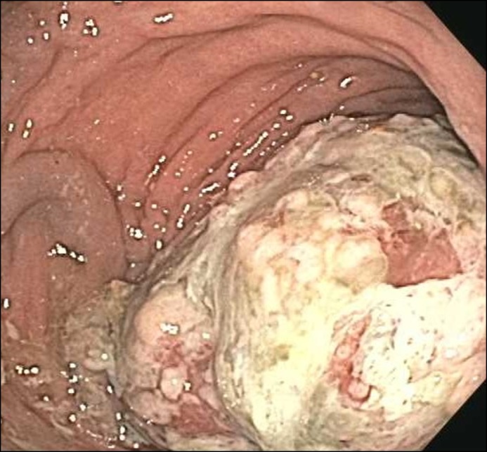
A 58-year-old woman with a history of chronic diarrhea and post-menopausal uterine bleeding underwent a routine colonoscopy for colon cancer screening, which demonstrated a large, nearly obstructing mass extending from the proximal rectum into the distal sigmoid colon (Figure 1). The colonoscope could be traversed proximally, and the remainder of the examination was normal. Biopsies from the lesion demonstrated granulation tissue and endometrial-type glands and stroma. Immunohistochemical stains showed CK7, PAX8, vimentin, estrogen and progesterone receptor–positive glands and stroma, consistent with endometriosis. No malignancy was identified even on deeper histologic sections. Due to the size of the mass, malignancy could not be excluded based on sampling. Her carcinoembryonic antigen (CEA) level was normal (3.9 ng/mL). A contrast-enhanced computed tomography (CT) scan of the abdomen and pelvis showed an enlarged heterogeneously enhancing uterus, rectosigmoid thickening with heterogeneous enhancement, and multiple similarly enhancing nodular soft tissues in the sigmoid mesentery and perirectal fat. CT scan of the chest did not show any evidence of metastatic disease.
Figure 1.

Colonoscopy showing a large mass partially occluding the lumen of the rectosigmoid colon.
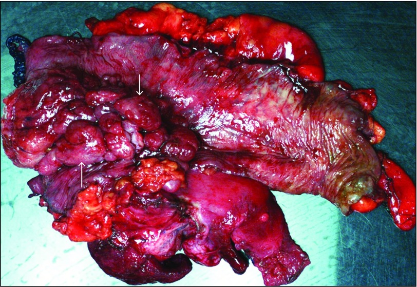
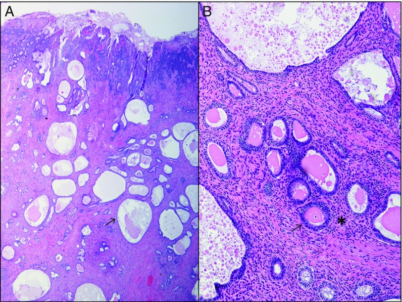
The patient underwent exploratory laparotomy with removal of the rectosigmoid mass, proctosigmoidectomy with low pelvic anastomosis and creation of proximal diverting loop ileostomy, total abdominal hysterectomy, and bilateral salpingo-oopherectomy. The pathology specimen revealed a 13 x 7 x 4-cm mass involving the colonic mucosa (Figure 2). Endometriosis was also found to involve the uterine cervix and tissue between the uterus and bowel. Seven pericolonic lymph nodes were biopsied and were negative for malignancy. Pathologic findings of surgical specimens showed endometrioid glands and stroma without atypia and foci of simple hyperplasia and mucinous metaplasia, consistent with a diagnosis of polypoid endometriosis without malignant transformation (Figure 3). Her postoperative course was notable for two separate hospital admissions for drainage from the abdominal midline vertical incision requiring wound vac placement and superficial cellulitis around ileostomy site secondary to the poor seal between the ostomy pouch and skin, but no other complications.
Figure 2.
A rectosigmoid colon mass (13.0 x 7.0 x 4.0 cm) showing polypoid endometriosis of the colonic mucosa.
Figure 3.
(A) Cystically dilated glands scattered beneath the colonic mucosa with focal crowding. (B) High-power view revealing benign-appearing endometrioid glands and stroma without atypia.
Polypoid endometriosis is a rare diagnosis that is challenging to diagnose on the basis of endoscopic findings and biopsy alone.1,2 Several nonspecific endoscopic findings have been described with this diagnosis, including polypoid lesions ranging from small sessile polyps to large pedunculated polyps in variable shades of white-gray, pink-red, or yellow-brown and are sometimes cystic or hemorrhagic.3 Lesions concerning for this process are often surgically resected given the difficulty to distinguish from malignancy and potential for malignant transformation.4,5
Disclosures
Author contributions: K. Han performed the literature review and wrote the manuscript. X. Li and R. Bhargava performed histopathological analysis and edited the manuscript. S. Taylor and K. Lee edited the manuscript. D. Yadav edited the manuscript and is the article guarantor.
Financial disclosure: None to report.
Informed consent was obtained for this case report.
References
- 1.Yamada Y, Miyamoto T, Horiuchi A, Ohya A, Shiozawa T. Polypoid endometriosis of the ovary mimicking ovarian carcinoma dissemination: A case report and literature review. J Obstet Gynaecol Res. 2014;40(5):1426–30. [DOI] [PubMed] [Google Scholar]
- 2.Jiang W, Roma A, Lai K, Carver P, Xiao S, Liu X. Endometriosis involving the mucosa of the intestinal tract: A clinicopathologic study of 15 cases. Mod Pathol. 2013;26:1270–8. [DOI] [PubMed] [Google Scholar]
- 3.Parker RL, Dadmanesh F, Young RH, Clement PB. Polypoid endometriosis: A clinicopathologic analysis of 24 cases and a review of the literature. Am J Surg Pathol. 2004;28:285–97. [DOI] [PubMed] [Google Scholar]
- 4.Takeuchi M, Matsuzaki K, Bando Y, Nishimura M, Yoneda A, Harada M. A case of polypoid endometriosis with malignant transformation. Abdom Radiol (NY). 2016;41(9):1699–702. [DOI] [PubMed] [Google Scholar]
- 5.Yantiss RK, Clement PB, Young RH. Neoplastic and pre-neoplastic changes in gastrointestinal endometriosis: A study of 17 cases. Am J Surg Pathol. 2000;24:513–24. [DOI] [PubMed] [Google Scholar]


